Review
. 2014 Sep 9:18:2331216514550621.
doi: 10.1177/2331216514550621.

Perceptual consequences of "hidden" hearing loss

Affiliations
Review

Perceptual consequences of "hidden" hearing loss

Christopher J Plack et al. Trends Hear. .

Abstract

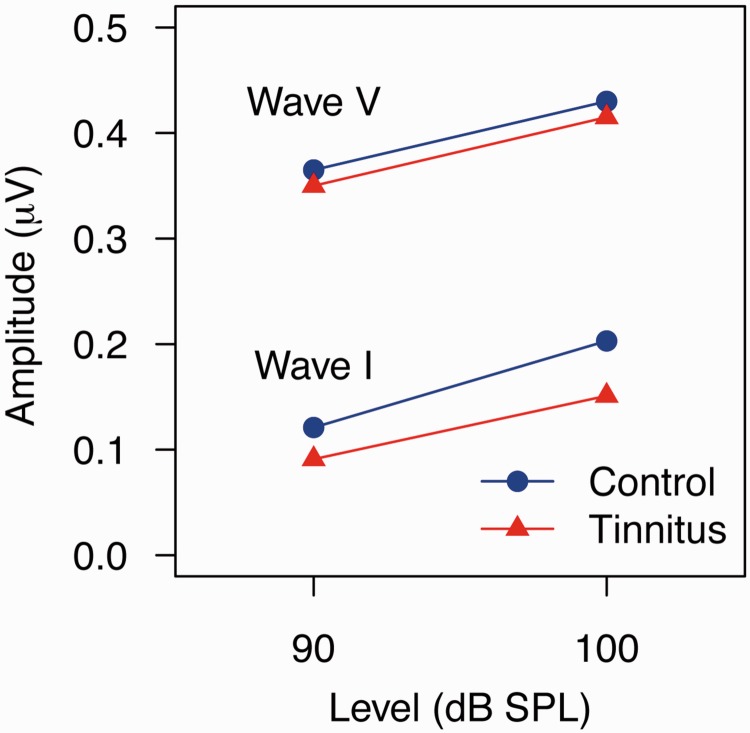
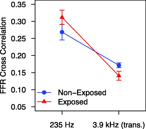
Dramatic results from recent animal experiments show that noise exposure can cause a selective loss of high-threshold auditory nerve fibers without affecting absolute sensitivity permanently. This cochlear neuropathy has been described as hidden hearing loss, as it is not thought to be detectable using standard measures of audiometric threshold. It is possible that hidden hearing loss is a common condition in humans and may underlie some of the perceptual deficits experienced by people with clinically normal hearing. There is some evidence that a history of noise exposure is associated with difficulties in speech discrimination and temporal processing, even in the absence of any audiometric loss. There is also evidence that the tinnitus experienced by listeners with clinically normal hearing is associated with cochlear neuropathy, as measured using Wave I of the auditory brainstem response. To date, however, there has been no direct link made between noise exposure, cochlear neuropathy, and perceptual difficulties. Animal experiments also reveal that the aging process itself, in the absence of significant noise exposure, is associated with loss of auditory nerve fibers. Evidence from human temporal bone studies and auditory brainstem response measures suggests that this form of hidden loss is common in humans and may have perceptual consequences, in particular, regarding the coding of the temporal aspects of sounds. Hidden hearing loss is potentially a major health issue, and investigations are ongoing to identify the causes and consequences of this troubling condition.

Keywords: aging; cochlear nerve; noise-induced hearing loss; sensorineural hearing loss; tinnitus.


Figures

Figure 1.
Figure 1.
An illustration of typical stimuli and recorded waveforms for two electrophysiological measures of auditory neural coding: the auditory brainstem response (ABR) and the frequency-following response (FFR). Note. Each trace represents the average of several thousand recordings. For the ABR, the waveform peaks reflect population neural activity at different stages in the auditory pathway following a brief click stimulus, from Wave I (auditory nerve) to Wave V (inferior colliculus). The FFR is a sustained response to a periodic stimulus, reflecting phase-locked neural activity in the rostral brainstem (region of the lateral lemniscus/inferior colliculus). The ABR and FFR can both be recorded by attaching electrodes to the scalp, for example, by using the differential response between electrodes on high forehead and mastoid.
Figure 2.
Figure 2.
The results of the study of Schaette and McAlpine (2011), showing the amplitudes of Wave I and Wave V of the ABR at two different click levels, for a group of tinnitus patients (triangles) and a group of audiogram-matched controls without tinnitus (circles). Note. Wave I amplitude is reduced in the tinnitus group relative to the control group, but there is no significant difference in Wave V amplitude between the groups. Data replotted from Schaette and McAlpine (2011).
Figure 3.
Figure 3.
A simulation of phase-locked auditory nerve activity in response to an amplitude modulated pure tone, illustrating the effects of loss of auditory nerve fibers on temporal coding. Note. The temporal fine structure of the stimulus waveform is shown in the top panel, and the envelope is shown as a dashed line. (a) Response of 10 auditory nerve fibers. (b) Response of three auditory nerve fibers. The summed response of 10 fibers (a, bottom) shows a clear representation of the envelope periodicity, and some representation of the fine structure. Temporal coding in the deafferented case (b, bottom) is sparser and less distinct.
Figure 4.
Figure 4.
Unpublished results from the conference presentation of Barker et al. (2014), showing FFR synchrony to a 235-Hz pure tone and to a 235-Hz tone transposed to 3.9 kHz (i.e., a 3.9-kHz pure-tone carrier amplitude modulated at 235 Hz), for groups of listeners with (triangles) and without (circles) a history of recreational noise exposure. Note. For each stimulus, the dependent variable was the coefficient of correlation between the FFR and a 235-Hz pure tone. The noise-exposed group show a (nonsignificantly) greater response to the 235-Hz pure tone than do the nonexposed group, but a significantly reduced response to the 235-Hz modulation component in the transposed tone, which is in the frequency region expected to be most susceptible to noise damage. Error bars show standard errors.
Figure 5.
Figure 5.
The results of the study of Marmel et al. (2013). Note. (a) Measure of FFR synchrony to a 660-Hz pure tone, plotted as a function of age. (b) Measure of FFR latency (group delay) as a function of age. Data replotted from Marmel et al. (2013).
Figure 6.
Figure 6.
Hypothetical causal links between noise-induced loss of AN fibers (cochlear neuropathy), deficits in the neural coding of temporal and intensity fluctuations, deficits in laboratory-based psychoacoustic tasks (lab. tasks), and deficits in real world hearing ability. Note. IPD stands for interaural phase difference, a temporal cue for localizing sounds based on direction-dependent differences in the arrival times of sounds at the two ears. Also shown are the hypothetical links between loss of AN fibers, central neural gain, and tinnitus and hyperacusis.

References

    1. Alvord L. S. (1983) Cochlear dysfunction in “normal-hearing” patients with history of noise exposure. Ear and Hearing 4: 247–250. - PubMed
    1. Baguley D. M. (2003) Hyperacusis. Journal of the Royal Society of Medicine 96: 582–585. - PMC - PubMed
    1. Barker, D., Hopkins, K., Baker, R., & Plack, C. J. (2014). Detecting the early effects of noise exposure. Paper presented at the Midwinter Meeting of Association for Research in Otolaryngology, San Diego, CA.
    1. Bharadwaj H. M., Verhulst S., Shaheen L., Liberman M. C., Shinn-Cunningham B. G. (2014) Cochlear neuropathy and the coding of supra-threshold sound. Frontiers in Systems Neuroscience 8: 26. - PMC - PubMed
    1. Borg E., Canlon B., Engstrom B. (1995) Noise-induced hearing loss. Literature review and experiments in rabbits. Morphological and electrophysiological features, exposure parameters and temporal factors, variability and interactions. Scandinavian Audiology Supplement 40: 1–147. - PubMed

Publication types

MeSH terms