. 2014 Oct 31;289(44):30459-30469.
doi: 10.1074/jbc.M114.591578. Epub 2014 Sep 9.

Genomic heat shock element sequences drive cooperative human heat shock factor 1 DNA binding and selectivity

Affiliations

Genomic heat shock element sequences drive cooperative human heat shock factor 1 DNA binding and selectivity

Alex M Jaeger et al. J Biol Chem. .

Abstract

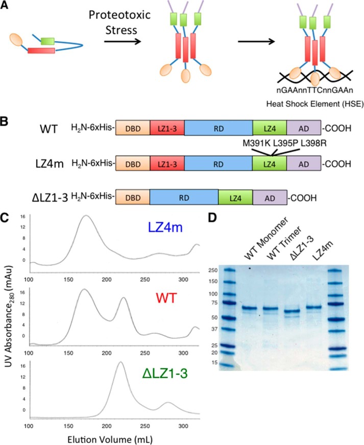
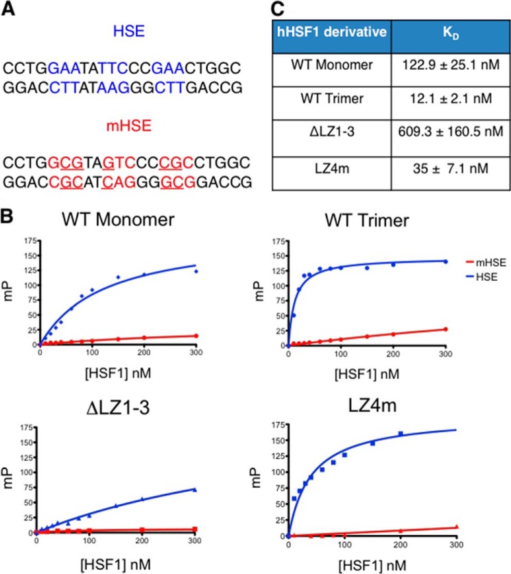
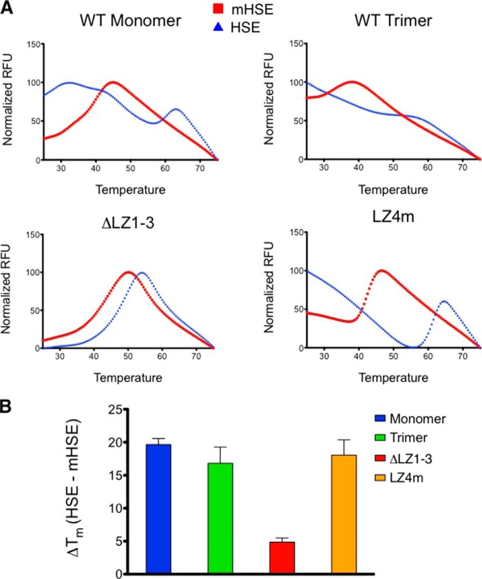
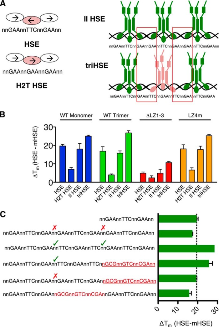
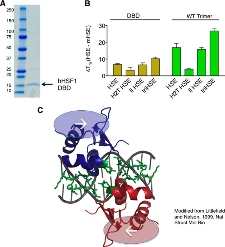
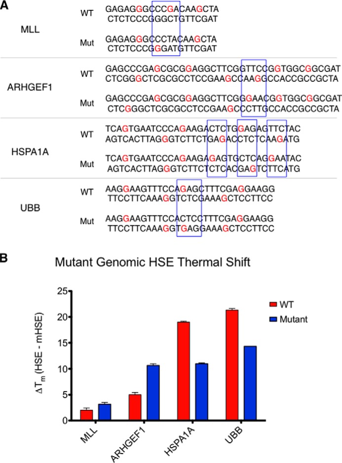
The heat shock transcription factor 1 (HSF1) activates expression of a variety of genes involved in cell survival, including protein chaperones, the protein degradation machinery, anti-apoptotic proteins, and transcription factors. Although HSF1 activation has been linked to amelioration of neurodegenerative disease, cancer cells exhibit a dependence on HSF1 for survival. Indeed, HSF1 drives a program of gene expression in cancer cells that is distinct from that activated in response to proteotoxic stress, and HSF1 DNA binding activity is elevated in cycling cells as compared with arrested cells. Active HSF1 homotrimerizes and binds to a DNA sequence consisting of inverted repeats of the pentameric sequence nGAAn, known as heat shock elements (HSEs). Recent comprehensive ChIP-seq experiments demonstrated that the architecture of HSEs is very diverse in the human genome, with deviations from the consensus sequence in the spacing, orientation, and extent of HSE repeats that could influence HSF1 DNA binding efficacy and the kinetics and magnitude of target gene expression. To understand the mechanisms that dictate binding specificity, HSF1 was purified as either a monomer or trimer and used to evaluate DNA-binding site preferences in vitro using fluorescence polarization and thermal denaturation profiling. These results were compared with quantitative chromatin immunoprecipitation assays in vivo. We demonstrate a role for specific orientations of extended HSE sequences in driving preferential HSF1 DNA binding to target loci in vivo. These studies provide a biochemical basis for understanding differential HSF1 target gene recognition and transcription in neurodegenerative disease and in cancer.

Keywords: Cancer; DNA-binding Protein; Gene Regulation; Neurodegeneration; Transcription Factor.


Figures

FIGURE 1.
FIGURE 1.
Expression and purification of human HSF1 derivatives. A, schematic of the current model of HSF1 activation. Upon stress, HSF1 transitions from a monomer to a trimer and binds HSEs throughout the genome. B, diagram of the constructs used for expressing HSF1 derivatives. WT human HSF1, HSF1 with mutations M391K, L395P, and L398R (LZ4m), and HSF1 with amino acids 138–198 deleted (ΔLZ1–3) were cloned into the pET15b E. coli expression vector with an amino-terminal His6 tag. C, gel filtration chromatography of HSF1 derivatives using Sephacryl s400 media. WT hHSF1 elutes as two distinct peaks, whereas ΔLZ1–3 and LZ4m elute as single peaks corresponding to HSF1 monomer and HSF1 trimer, respectively. Each peak was isolated and concentrated to 1 mg/ml. D, SDS-PAGE analysis of 2 μg of the four species of purified HSF1.
FIGURE 2.
FIGURE 2.
Fluorescence polarization analysis of HSF1-HSE binding correlates with multimeric state of HSF1 derivatives. A, sequences used for FP analysis. dsDNA containing the inverted nGAAn repeats (HSE) was used for specific DNA binding. A dsDNA with specific mutations disrupting the nGAAn repeat (mHSE) was used as a negative control. B, representative binding curves generated from FP experiments for the four HSF1 derivatives. An increase in millipolarization units (MP) is indicative of direct binding to the fluorescently labeled HSE probe. The HSF1 variants saturate the HSE probe with different binding kinetics. C, Kd values calculated from a hyperboloidal curve fit for the four HSF1 derivatives.
FIGURE 3.
FIGURE 3.
Thermal denaturation profiling of HSF1 derivatives. A, representative melting curves for all derivatives in the presence of nonspecific (mHSE) or specific (HSE) DNA. WT monomer, WT trimer, and LZ4m demonstrate enhanced thermal stability in the presence of HSE DNA. ΔLZ1–3 is also stabilized but to a much lesser extent than the other HSF1 derivatives. B, thermal shifts calculated from the melting curves. The first derivative of the melting curve was calculated, and the highest derivative value was taken as the melting temperature. Melting temperatures of HSF1 in the presence of the mHSE were subtracted from melting temperatures for the HSE curves for each HSF1 derivative to provide a thermal shift (ΔTm). WT monomer, WT trimer, and LZ4m are stabilized by ∼20 °C, whereas ΔLZ1–3 stability is shifted by 4 °C.
FIGURE 4.
FIGURE 4.
Thermal denaturation profiling of HSE variants identifies a role for extended HSEs. A, diagram of the HSE variants used. HSE is the same as used in Fig. 3. Head to tail (H2T) HSE is generated by inverting the middle nGAAn repeat of the HSE sequence. This disrupts the typical head to head interface of the DNA binding domain while bound to DNA. The II HSE is constructed by placing three HSE sites in tandem with an orientation that provides a head to tail interaction between trimers (red boxes). The triple HSE (triHSE) is three HSEs oriented so that head to head or tail to tail interactions (red boxes) are present between trimers. B, ΔTm values in the presence of HSE variants. C, triHSE variants with one HSE disrupted by an mHSE. A triHSE with two trimer binding sites oriented in a cooperative fashion imparts a thermal shift between a single HSE and triHSE, whereas a triHSE with two trimer binding sites in noncooperative orientation or separated by a mHSE do not demonstrate an increased thermal stability.
FIGURE 5.
FIGURE 5.
HSF1 DNA-binding domain participates in cooperative HSE binding. A, SDS-PAGE analyses of the purified HSF1 DBD with molecular weight indicated. B, DBD and WT trimer thermal shifts in the presence of HSE variants. Although the magnitude of the thermal shift is different between the two HSF1 derivatives, a similar relationship is observed between the different HSEs for the two derivatives. C, representation of the K. lactis DBD crystal structure (25) modeled in PyMOL. The dimer interface (green) observed in the crystal structure is predicted to be present between trimers in cooperative HSEs but not in noncooperative HSEs, suggesting that this interface may contribute to cooperative binding to extended HSEs in the proper orientation.
FIGURE 6.
FIGURE 6.
HSF1 binding to genomic HSEs in vitro by thermal denaturation correlates with binding in vivo. A, HSF1 thermal shifts elicited by genomic HSEs. The magnitude of thermal shift correlates with the cooperative nature of the HSE. B, chromatin immunoprecipitation analysis of endogenous human HEK293T cell HSF1 binding to genomic sequences tested in B. A strong correlation can be drawn between in vitro HSE binding as assessed by thermal shift and in vivo binding measured by quantitative ChIP.
FIGURE 7.
FIGURE 7.
Directed mutation of cooperativity within genomic HSE sequences results in changes in HSF1 DNA binding affinity. A, sequences of “wild type” (WT) and mutant (Mut) genomic HSEs within the MLL, ARHGEF1, HSPA1A, and UBB genes. Subtle mutations within the MLL and ARHGEF1 HSEs foster increased cooperativity, whereas mutations within the HSPA1A and UBB sequence disrupt cooperative interactions between trimers. B, thermal shifts elicited by the genomic HSE variants. MLL and ARHGEF1 HSE mutants exhibit higher affinity binding, whereas the HSPA1A and UBB HSE mutants exhibit lower binding affinity for HSF1. All mutants demonstrated significantly different melting profiles as measured by one-way analysis of variance followed by Newman-Keuls multiple comparison test.

References

    1. Bukau B., Weissman J., Horwich A. (2006) Molecular chaperones and protein quality control. Cell 125, 443–451 - PubMed
    1. Neef D. W., Jaeger A. M., Thiele D. J. (2011) Heat shock transcription factor 1 as a therapeutic target in neurodegenerative diseases. Nat. Rev. Drug Discov. 10, 930–944 - PMC - PubMed
    1. Akerfelt M., Morimoto R. I., Sistonen L. (2010) Heat shock factors: integrators of cell stress, development, and lifespan. Nat. Rev. Mol. Cell Biol. 11, 545–555 - PMC - PubMed
    1. Glover-Cutter K. M., Lin S., Blackwell T. K. (2013) Integration of the unfolded protein and oxidative stress responses through SKN-1/Nrf. PLoS Genet. 9, e1003701. - PMC - PubMed
    1. Morimoto R. I. (2008) Proteotoxic stress and inducible chaperone networks in neurodegenerative disease and aging. Genes Dev. 22, 1427–1438 - PMC - PubMed

Publication types