. 2014 Nov;184(11):3142-53.
doi: 10.1016/j.ajpath.2014.07.017. Epub 2014 Sep 7.

The membrane attack complex in aging human choriocapillaris: relationship to macular degeneration and choroidal thinning

Affiliations

The membrane attack complex in aging human choriocapillaris: relationship to macular degeneration and choroidal thinning

Robert F Mullins et al. Am J Pathol. 2014 Nov.

Abstract

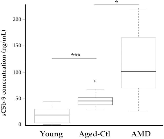
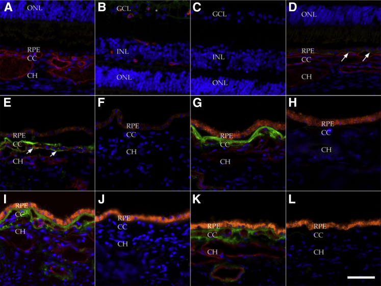
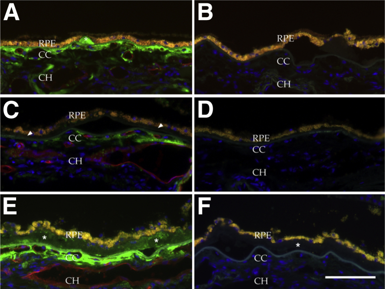
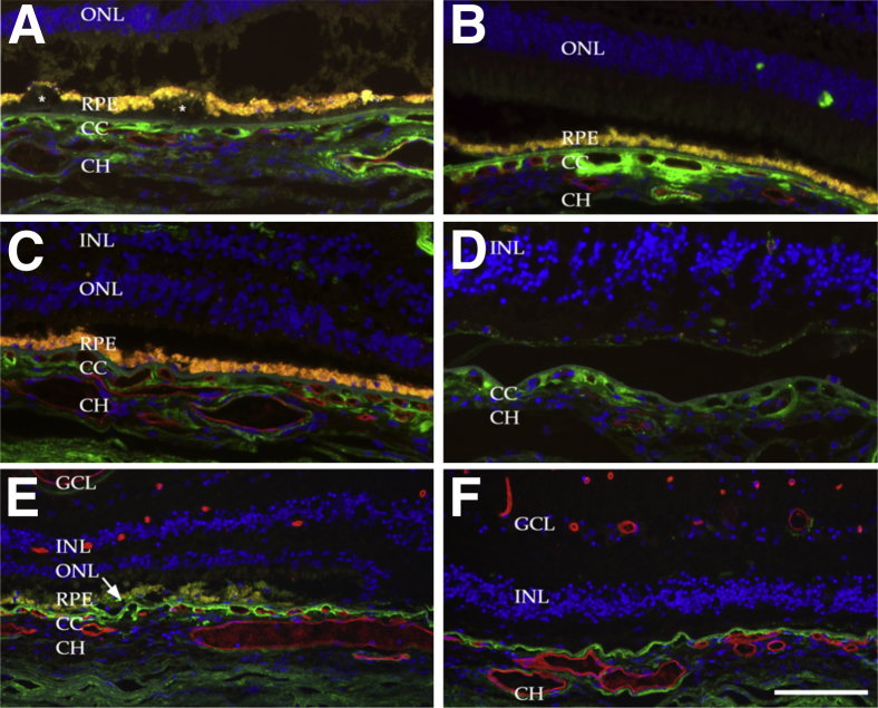
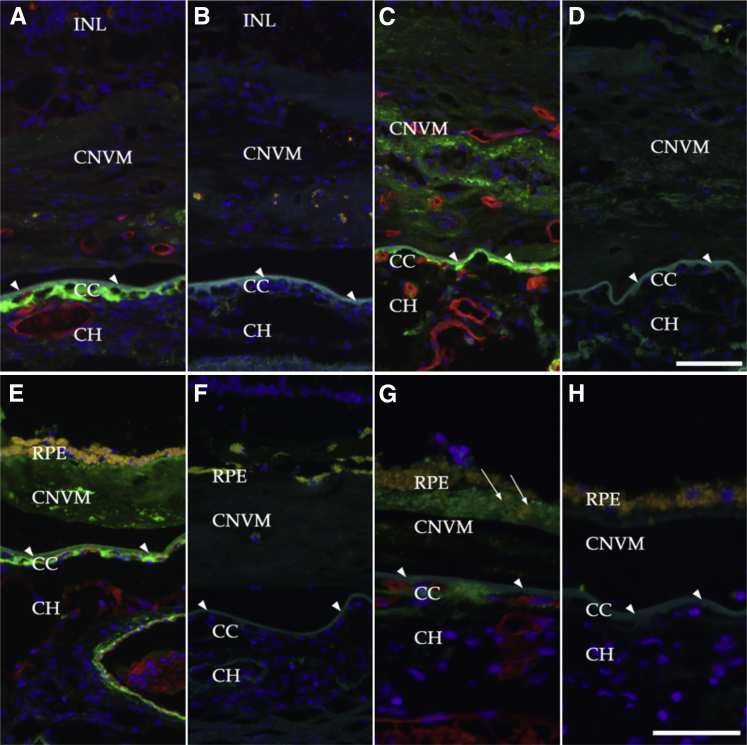
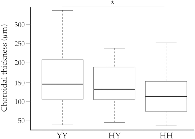
Age-related macular degeneration (AMD) is a common disease that can result in severe visual impairment. Abnormal regulation of the complement system has been implicated in its pathogenesis, and CFH polymorphisms contribute substantially to risk. How these polymorphisms exert their effects is poorly understood. We performed enzyme-linked immunosorbent assay (ELISA) analysis on young, aged, and AMD choroids to determine the abundance of the membrane attack complex (MAC) and performed immunofluorescence studies on eyes from 117 donors to evaluate the MAC in aging, early AMD, and advanced AMD. Morphometric studies were performed on eyes with high- or low-risk CFH genotypes. ELISA confirmed that MAC increases significantly with aging and with AMD. MAC was localized to Bruch's membrane and the choriocapillaris and was detectable at low levels as early as 5 years of age. Hard drusen were labeled with anti-MAC antibody, but large or confluent drusen and basal deposits were generally unlabeled. Labeling of retinal pigment epithelium was observed in some cases of advanced AMD, but not in early disease. Eyes homozygous for the high-risk CFH genotype had thinner choroids than low-risk homozygotes (P < 0.05). These findings suggest that increased complement activation in AMD and in high-risk genotypes can lead to loss of endothelial cells in early AMD. Treatments to protect the choriocapillaris in early AMD are needed.


Figures

Figure 1
Figure 1
Enzyme-linked immunosorbent assay analysis of soluble C5b-9 MAC in young eyes, aged eyes without AMD (control), and eyes with atrophic AMD. Increased MAC levels were related to increased age (∗∗∗P < 0.001) and to diagnosis of AMD (P < 0.05). Data are expressed as box-and-whisker plots, indicating median, interquartile range, and minimum and maximum values; any outliers are indicated by individual symbols. n = 10 per group. AMD, age-related macular degeneration; Ctl, control; MAC, membrane attack complex.
Figure 2
Figure 2
Localization of the membrane attack complex (MAC) in young (<50 years) and aged donor eyes without AMD: newborn (A–C), 16 months (D), 5 years (E and F), 32 years (G and H), 41 years (I and J), and 62 years (K and L). Sections were dual-labeled with anti-C5b-9 MAC antibody (green) and UEA-I lectin (red) (A, B, D, E, G, I, and K); the primary antibody and lectin were omitted for negative control (C, F, H, J, and L). Orange fluorescence of the RPE (G–L) indicates age-related lipofuscin accumulation. A–C: Eyes from a newborn infant showed no MAC labeling in Bruch’s membrane or choroid (A). Minor immunoreactivity was observed in the ganglion cell layer (B, asterisks; C, negative control) of this newborn, but in none of the older specimens. D: At 16 months of age, very minor labeling was observed as puncta (arrows) between the RPE and choriocapillaris. E: In a 5-year-old donor heterozygous for the Y402H variant, considerable MAC labeling (arrows) in Bruch’s membrane extended around the choriocapillaris. G–L: More robust labeling was observed in older eyes. Note the lack of choriocapillaris labeling and the constitutive RPE autofluorescence in the negative controls (H, J, and L). All images were processed identically. Scale bar = 50 μm. CC, choriocapillaris; CH, outer choroid; GCL, ganglion cell layer; INL, inner nuclear layer; ONL, outer nuclear layer; RPE, retinal pigment epithelium.
Figure 3
Figure 3
Localization of the MAC in eyes with early AMD. A: MAC was localized to solitary, hard drusen (asterisk). C: By contrast, basal deposits characteristic of early AMD were generally unreactive with anti-MAC antibody (arrowheads). E: Modest punctate immunoreactivity was observed where drusen were confluent (asterisks). Note immunoreactive extracellular material extending into the outer choroid. B, D, and F: Secondary antibody controls from adjacent sections. Scale bar = 100 μm.
Figure 4
Figure 4
Localization of the MAC in the choriocapillaris of eyes with geographic atrophy. The choroid is thinner in eyes with advanced dry AMD than in control eyes. Shown are areas outside (A–C), within (D and F), and at the junction (E) of the central atrophy. A: Drusen deposits (asterisks), extensive MAC in the choriocapillaris layer, and vascular atrophy with loss of endothelium (ghost vessels) in an 83-year-old donor eye. B: Choriocapillaris atrophy, with nonreactive basal deposits, in an 82-year-old donor eye. C: An area of atrophy in an 89-year-old donor eye, with some thinning and attenuation of the ONL and shortening of the inner–outer segment. D: An area of central atrophy, with loss of the RPE, in an 87-year-old donor eye. Note the modest amount of RPE lipofuscin in a gliotic scar. MAC immunoreactivity was still present in the largely atrophied choriocapillaris. E and F: The atrophic interface (E) and the central atrophic zone (F) of an eye from a 68-year-old donor homozygous for the high-risk Y402H allele. Note localization of MAC to RPE cells near the interface (E, arrow) and persistence of MAC in the degenerated choroid in the area of central atrophy. Scale bar = 100 μm. CC, choriocapillaris; CH, outer choroid; GCL, ganglion cell layer; INL, inner nuclear layer; ONL, outer nuclear layer; RPE, retinal pigment epithelium.
Figure 5
Figure 5
Localization of MAC in eyes with choroidal neovascularization under dual labeling with anti-C9 neoepitope antibody (green) and the vascular-labeling lectin UEA-I (red). Sections were incubated with primary and secondary antibody (A, C, E, and G); adjacent unlabeled sections served as negative controls (B, D, F, and H). Bruch’s membrane is indicated by arrowheads. A and C: MAC persisted in domains surrounding the choriocapillaris even after extensive atrophy. MAC was also present in some choroidal neovascular membranes in association with dedifferentiated RPE cells (C, arrows). E: Punctate labeling could also be observed in subretinal fibrosis and beneath the RPE that resided on the surface of these fibrotic membranes. G: In an eye with type I or occult CNVM, MAC labeling of the RPE was robust (arrows). Scale bars: 50 μm (G and H); 75 μm (A–F). CC, choriocapillaris; CH, outer choroid; CNV, choroidal neovascularization; CNVM, choroidal neovascular membrane; INL, inner nuclear layer; RPE, retinal pigment epithelium.
Figure 6
Figure 6
Choroidal thickness in donor eyes of the three Y402H CFH genotypes. Measurements of choroidal thickness were performed as described previously, and genotypes were determined for the Y402H allele (rs1061170). Eyes from donors homozygous for the high-risk H allele had thinner choroids than eyes from donors heterozygous or homozygous for the low-risk Y allele. Eyes homozygous for the H allele had significantly thinner choroids than those homozygous for the Y allele (P < 0.05), and there was a significant association (P = 0.019) between choroidal thickness and the number of copies of the H allele. Data are expressed as box-and-whisker plots, indicating median, interquartile range, and minimum and maximum values. n = 43 (YY); n = 32 (HY); n = 25 (HH). P < 0.05.

References

    1. Vannewkirk M.R., Nanjan M.B., Wang J.J., Mitchell P., Taylor H.R., McCarty C.A. The prevalence of age-related maculopathy: the Visual Impairment Project. Ophthalmology. 2000;107:1593–1600. - PubMed
    1. Friedman D.S., O’Colmain B.J., Muñoz B., Tomany S.C., McCarty C., de Jong P.T.V.M., Nemesure B., Mitchell P., Kempen J., Eye Diseases Prevalence Research Group Prevalence of age-related macular degeneration in the United States. Arch Ophthalmol. 2004;122:564–572. [Erratum appeared in Arch Ophthalmol 2011, 129:1188] - PubMed
    1. Bird A.C., Bressler N.M., Bressler S.B., Chisholm I.H., Coscas G., Davis M.D., de Jong P.T.V.M., Klaver C.C.W., Klein B.E.K., Klein R., Mitchell P., Sarks J.P., Sarks S.H., Soubrane G., Taylor H.R., Vingerling J.R., International ARM Epidemiological Study Group An international classification and grading system for age-related maculopathy and age-related macular degeneration. Surv Ophthalmol. 1995;39:367–374. - PubMed
    1. Khandhadia S., Cipriani V., Yates J.R.W., Lotery A.J. Age-related macular degeneration and the complement system. Immunobiology. 2012;217:127–146. - PubMed
    1. Haines J.L., Hauser M.A., Schmidt S., Scott W.K., Olson L.M., Gallins P., Spencer K.L., Kwan S.Y., Noureddine M., Gilbert J.R., Schnetz-Boutaud N., Agarwal A., Postel E.A., Pericak-Vance M.A. Complement factor H variant increases the risk of age-related macular degeneration. Science. 2005;308:419–421. - PubMed

Publication types

MeSH terms

Substances