. 2014 Nov;184(11):2965-75.
doi: 10.1016/j.ajpath.2014.07.014. Epub 2014 Sep 6.

The microbiota protects against ischemia/reperfusion-induced intestinal injury through nucleotide-binding oligomerization domain-containing protein 2 (NOD2) signaling

Affiliations

The microbiota protects against ischemia/reperfusion-induced intestinal injury through nucleotide-binding oligomerization domain-containing protein 2 (NOD2) signaling

Ernesto Perez-Chanona et al. Am J Pathol. 2014 Nov.

Abstract

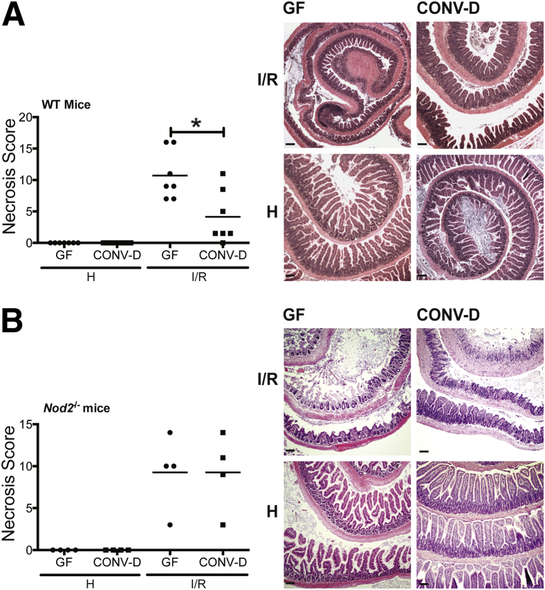
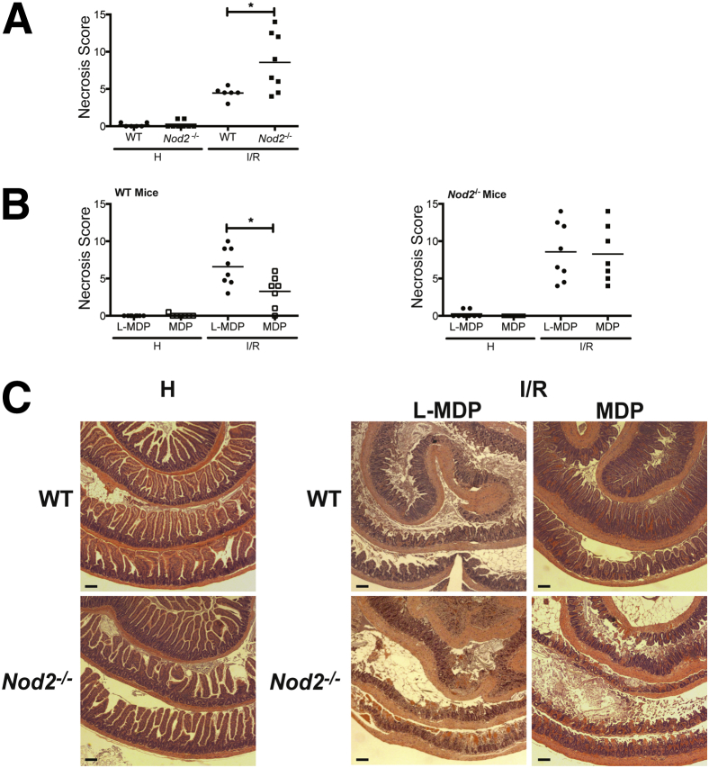
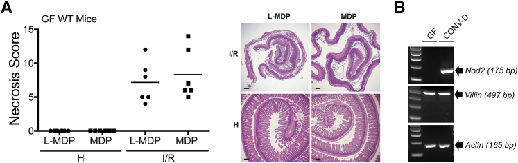
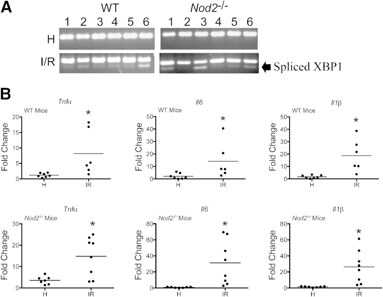
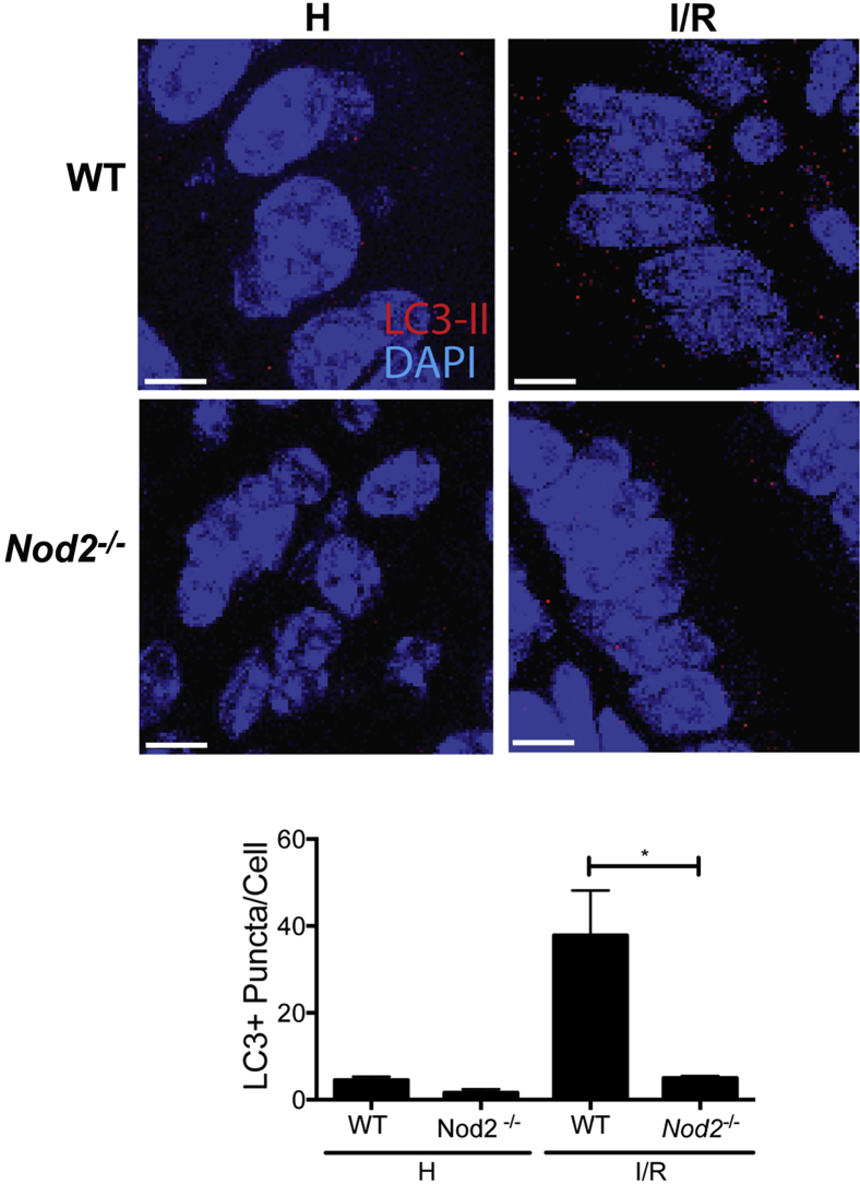
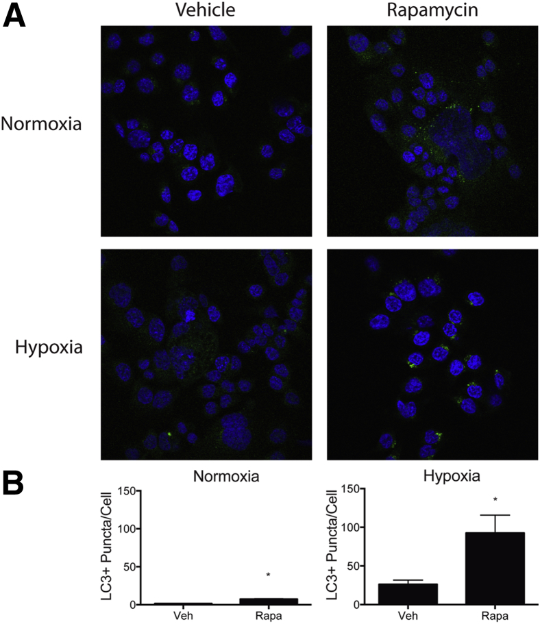
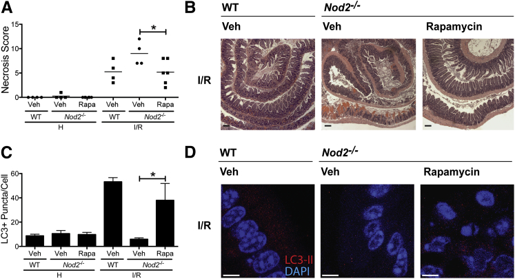
Nucleotide-binding oligomerization domain-containing protein 2 (NOD2), an intracellular pattern recognition receptor, induces autophagy on detection of muramyl dipeptide (MDP), a component of microbial cell walls. The role of bacteria and NOD2 signaling toward ischemia/reperfusion (I/R)-induced intestinal injury response is unknown. Herein, we report that I/R-induced intestinal injury in germ-free (GF) C57BL/6 wild-type (WT) mice is worse than in conventionally derived mice. More important, microbiota-mediated protection against I/R-induced intestinal injury is abrogated in conventionally derived Nod2(-/-) mice and GF Nod2(-/-) mice. Also, WT mice raised in specific pathogen-free (SPF) conditions fared better against I/R-induced injury than SPF Nod2(-/-) mice. Moreover, SPF WT mice i.p. administered 10 mg/kg MDP were protected against injury compared with mice administered the inactive enantiomer, l-MDP, an effect lost in Nod2(-/-) mice. However, MDP administration failed to protect GF mice from I/R-induced intestinal injury compared with control, a phenomenon correlating with undetectable Nod2 mRNA level in the epithelium of GF mice. More important, the autophagy-inducer rapamycin protected Nod2(-/-) mice against I/R-induced injury and increased the levels of LC3(+) puncta in injured tissue of Nod2(-/-) mice. These findings demonstrate that NOD2 protects against I/R and promotes wound healing, likely through the induction of the autophagy response.


Figures

Figure 1
Figure 1
Microbial signaling protects against I/R-induced injury. Cohorts of GF, WT mice and GF, Nod2−/− mice were split into two groups. One group remained in germ free (GF) conditions, whereas the other was CONV-D with commensal microbes in a SPF facility. Mice were subjected to 60 minutes of ischemia, followed by 3 hours of reperfusion. Necrosis was assessed using an established necrosis scoring system. Histological intestinal damage scores of individual mice are depicted (means ± SEM), and representative images of H&E-stained ileal sections of WT (A) and Nod2−/− (B) mice are shown. Results are representative of two independent experiments (n ≥ 4 per group). P < 0.05. Scale bar = 100 μm (A and B). H, healthy; I/R, 1 hour ischemia, followed by 3 hours’ reperfusion.
Figure 2
Figure 2
MDP signaling protects against I/R-induced injury. WT and Nod2−/− mice were subjected to ileal ischemia for 1 hour, followed by 1.5 hours of reperfusion. NOD2 ligand, 1 mg/kg MDP, or 1 mg/kg of its enantiomer, l-MDP, was injected i.p. in WT mice 24 hours before I/R exposure. Healthy and injured tissues were collected, Swiss rolled, and stained with H&E. Necrosis was assessed using an established necrosis scoring system. A and B: Histological intestinal damage scores of individual mice are depicted. C: Representative images of H&E-stained ileal sections. Results are representative of three independent experiments (n ≥ 4 per group). Data are given as means ± SEM (A and B). P < 0.05. Scale bar = 100 μm (C). H, healthy; I/R, 1 hour ischemia, followed by 1.5 hours’ reperfusion.
Figure 3
Figure 3
Microbial induction of NOD2 expression in IECs is necessary for protection against injury. A: Germ free (GF), WT mice were subjected to ileal ischemia for 1 hour, and followed by 3 hours of reperfusion. NOD2 ligand, 1 mg/kg MDP, or 1 mg/kg of its enantiomer, l-MDP, was injected i.p. in mice 24 hours before I/R exposure. Healthy and injured tissue was collected, Swiss rolled, and stained with H&E. Necrosis was assessed using an established necrosis scoring system. Histological intestinal damage scores of individual mice are depicted (left), and representative images of H&E-stained ileal sections are shown (right). B: IECs were isolated from GF and CONV-D WT mice, and Nod2 mRNA level was assessed using RT-PCR. Actin and Villin mRNAs were amplified as loading and isolation efficacy, respectively. Scale bar = 100 μm (B). H, healthy; I, ischemia only; I/R, 1 hour ischemia, followed by 3 hours’ reperfusion.
Figure 4
Figure 4
I/R-induced injury induces ER stress in WT and Nod2−/− mice. WT and Nod2−/− mice were subjected to I/R-induced injury. A: RT-PCR of ER-stress response marker, XBP1, was performed and resolved on a 3% agarose gel. B: Il1b, Il6, and Tnfα mRNAs from healthy and injured ileal tissues were determined using ABI Prism 7900HT (Life Technologies, Grand Island, NY). Data were processed using the ΔΔCT method, normalized to β-actin, and set relative to healthy tissue. Results are representative of three independent experiments. P < 0.05 (n ≥ 4 per group). H, healthy; I/R, 1 hour ischemia, followed by 1.5 hours’ reperfusion.
Figure 5
Figure 5
I/R-induced injury induces autophagy through NOD2. Immunofluorescence staining of LC3 was performed on paraffin-embedded healthy and injured ileal tissue from WT and Nod2−/− mice. NOD2 ligand, 1 mg/kg MDP, or 1 mg/kg of its enantiomer, l-MDP, was injected i.p. in mice 24 hours before I/R exposure. The average number of LC3+ puncta per cell in the injured and healthy portions of the terminal ileum of untreated WT and Nod2−/− mice and corresponding representative images of the intestinal epithelium of mice. P < 0.05 (results are representative of three fields of view per sample). Scale bar = 5 μm. H, healthy; I/R, 1 hour ischemia, followed by 1.5 hours’ reperfusion.
Figure 6
Figure 6
Rapamycin (Rapa) enhances hypoxia-induced autophagy in human colon colorectal carcinoma HCT116 cells. HCT116 cells were treated with 10 μg/mL pepstatin A and 10 μg/mL E-64-d 1 hour before exposure to 1 hour of normoxia or hypoxia (1% O2, Coy Labs hypoxic glove box) and stimulated by 5 μg/mL Rapa. LC3+ puncta (green fluorescent protein) and cells (DAPI) were counted using ImageJ software. A: Representative images of LC3+ puncta in HCT116 cells stimulated with Rapa in normoxic and hypoxic conditions. B: The average number of puncta per cell of cells stimulated with Rapa in normoxic and hypoxic conditions. Results are representative of three independent experiments. P < 0.05. Veh, vehicle.
Figure 7
Figure 7
Rapamycin (Rapa)–induced autophagy protects Nod2−/− mice against I/R-induced injury. WT and Nod2−/− mice were subjected to ileal ischemia, followed by 1.5 hours of reperfusion. Autophagy-inducer, Rapa (3 mg/kg), or its vehicle (Veh), DMSO, was injected i.p. in Nod2−/− 1 hour before I/R exposure. Healthy and injured tissues were collected, Swiss rolled, and stained with H&E. Necrosis was assessed using an established necrosis scoring system. A: Histological intestinal damage scores of individual. B: Representative images of H&E-stained ileal sections. Results are representative of two independent experiments (n ≥ 4 per group). C and D: The average number of LC3+ puncta per cell and representative images of the intestinal epithelium of mice administered either Rapa or Veh. Results are representative of three independent fields of view per sample. Data are given as means ± SEM (A). P < 0.05. Scale bars: 100 μm (B); 5 μm (C and D). H, healthy; I/R, 1 hour ischemia, followed by 1.5 hours of reperfusion; Veh, vehicle.

References

    1. Abreu M.T. Toll-like receptor signalling in the intestinal epithelium: how bacterial recognition shapes intestinal function. Nat Rev Immunol. 2010;10:131–144. - PubMed
    1. Prescott D., Lee J., Philpott D.J. An epithelial armamentarium to sense the microbiota. Semin Immunol. 2013;25:323–333. - PubMed
    1. Karrasch T., Jobin C. Wound healing responses at the gastrointestinal epithelium: a close look at novel regulatory factors and investigative approaches. Z Gastroenterol. 2009;47:1221–1229. - PubMed
    1. Eltzschig H.K., Eckle T. Ischemia and reperfusion—from mechanism to translation. Nat Med. 2011;17:1391–1401. - PMC - PubMed
    1. Farber A., Connors J.P., Friedlander R.M., Wagner R.J., Powell R.J., Cronenwett J.L. A specific inhibitor of apoptosis decreases tissue injury after intestinal ischemia-reperfusion in mice. J Vasc Surg. 1999;30:752–760. - PubMed

Publication types

Substances

LinkOut - more resources