Review

Proposed guidelines for the diagnosis and management of methylmalonic and propionic acidemia

Matthias R Baumgartner et al. Orphanet J Rare Dis. .

Abstract

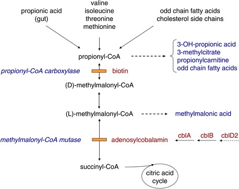
Methylmalonic and propionic acidemia (MMA/PA) are inborn errors of metabolism characterized by accumulation of propionic acid and/or methylmalonic acid due to deficiency of methylmalonyl-CoA mutase (MUT) or propionyl-CoA carboxylase (PCC). MMA has an estimated incidence of ~ 1: 50,000 and PA of ~ 1:100'000 -150,000. Patients present either shortly after birth with acute deterioration, metabolic acidosis and hyperammonemia or later at any age with a more heterogeneous clinical picture, leading to early death or to severe neurological handicap in many survivors. Mental outcome tends to be worse in PA and late complications include chronic kidney disease almost exclusively in MMA and cardiomyopathy mainly in PA. Except for vitamin B12 responsive forms of MMA the outcome remains poor despite the existence of apparently effective therapy with a low protein diet and carnitine. This may be related to under recognition and delayed diagnosis due to nonspecific clinical presentation and insufficient awareness of health care professionals because of disease rarity.


Figures

Figure 1
Figure 1
Metabolic interrelationship of MMA and PA.
Figure 2
Figure 2
Neuroimaging finding in MMA: Male child with MMA (cblA defect). An MRI study was obtained at the age of 6 months because of irritability, feeding difficulties, developmental delay, encephalopathy and metabolic decompensation. The caudate and lentiform nuclei are swollen and hyperintense on axial T2-weighted (W) turbo spin-echo (SE) image (A) with restricted diffusion suggested by hyperintensity on trace diffusion-weighted imaging (DWI) (B) and low signals on ADC map (C). Proton MR spectroscopy (TE:135ms) performed from the lesions reveals decreased N-acetyl-aspartate (NAA), increased choline (Cho) and presence of lactate (D).
Figure 3
Figure 3
Neuroimaging finding in MMA: Male child with MMA (cblA defect). Follow-up MR imaging 6 months after acute deterioration shows residual T2 hyperintensity in the caudate nuclei and necrosis in the lentiform nuclei with reversal of abnormalities in the globus pallidi (A). There is CSF-like unrestricted diffusion in the lentiform nuclei on DWI (B) and ADC map (C) and elevated Cho on MR spectroscopy (D).
Figure 4
Figure 4
Neuroimaging finding in PA: Male child with PA, symptomatic from day 4 of life, diagnosed at the age of 4 months. An MRI was obtained at the age of 5 years when he developed acute encephalopathy. Cerebral cortices especially of the temporal and occipital lobes and the basal ganglia (caudate and lentiform nuclei) are swollen and mildly hyperintense on axial T2W TSE (A). Hyperintensity on trace DWI (B) and low intensity on ADC maps (C) suggestive of restricted diffusion are seen in those affected regions.
Figure 5
Figure 5
Neuroimaging finding in PA: Male child with PA, symptomatic from day 4 of life, diagnosed at the age of 4 months. A week later a repeat MR study showed more intense T2 signal changes in the involved regions, more prominent in the putamina (A). Although a higher signal intensity of the cortices and basal ganglia on trace DWI (B), with disappearance of low signal of the cortices and presence of higher signal intensity on ADC maps (C) there’s pseudonormalization of diffusion restriction.

References

    1. Fowler B, Leonard JV, Baumgartner MR. Causes of and diagnostic approach to methylmalonic acidurias. J Inherit Metab Dis. 2008;31:350–360. doi: 10.1007/s10545-008-0839-4. - DOI - PubMed
    1. Coelho D, Suormala T, Stucki M, Lerner-Ellis JP, Rosenblatt DS, Newbold RF, Baumgartner MR, Fowler B. Gene identification for the cblD defect of vitamin B12 metabolism. N Engl J Med. 2008;358:1454–1464. doi: 10.1056/NEJMoa072200. - DOI - PubMed
    1. Coelho D, Kim JC, Miousse IR, Fung S, du Moulin M, Buers I, Suormala T, Burda P, Frapolli M, Stucki M, Nürnberg P, Thiele H, Robenek H, Höhne W, Longo N, Pasquali M, Mengel E, Watkins D, Shoubridge EA, Majewski J, Rosenblatt DS, Fowler B, Rutsch F, Baumgartner MR. Mutations in ABCD4 cause a new inborn error of vitamin B12 metabolism. Nat Genet. 2012;44:1152–1155. doi: 10.1038/ng.2386. - DOI - PubMed
    1. Deodato F, Boenzi S, Santorelli FM, Dionisi-Vici C. Methylmalonic and propionic aciduria. Am J Med Genet C Semin Med Genet. 2006;142C:104–112. doi: 10.1002/ajmg.c.30090. - DOI - PubMed
    1. Manoli I, Venditti C. Methylmalonic Acidemia. In: Pagon RA, Adam MP, Ardinger HH, Bird TD, Dolan CR, Fong CT, Smith RJH, Stephens K, editors. GeneReviews®. Seattle: University of Washington; 2005.

Publication types

MeSH terms

Supplementary concepts