. 2014 Dec;65(22):6487-98.
doi: 10.1093/jxb/eru366. Epub 2014 Sep 9.

Expression patterns of flagellin sensing 2 map to bacterial entry sites in plant shoots and roots

Affiliations

Expression patterns of flagellin sensing 2 map to bacterial entry sites in plant shoots and roots

Martina Beck et al. J Exp Bot. 2014 Dec.

Abstract

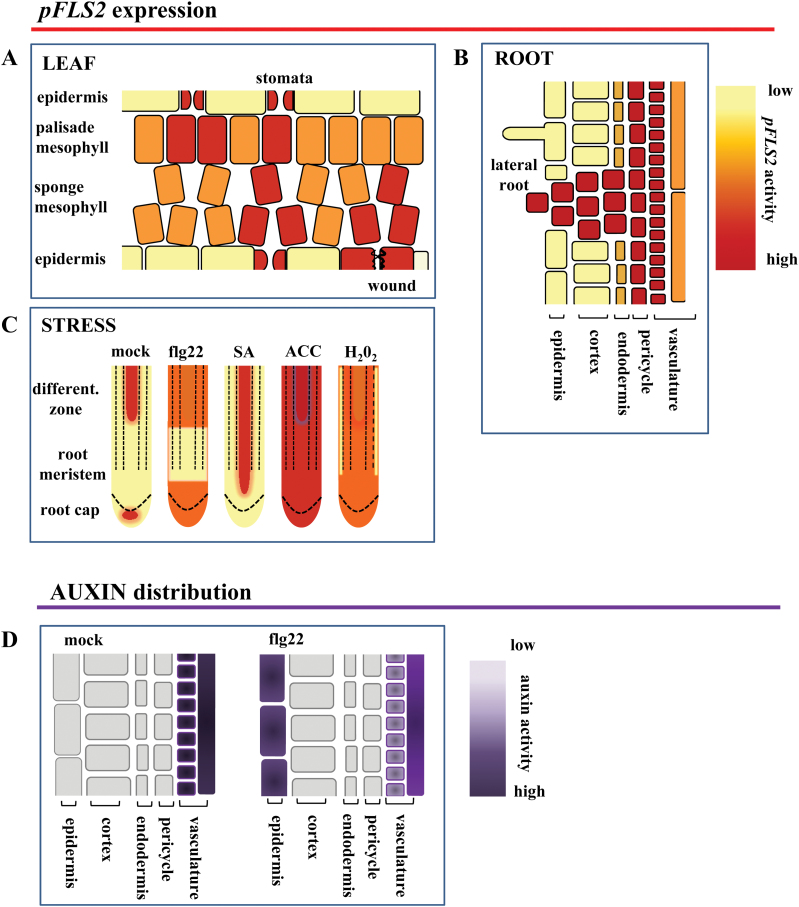
Pathogens can colonize all plant organs and tissues. To prevent this, each cell must be capable of autonomously triggering defence. Therefore, it is generally assumed that primary sensors of the immune system are constitutively present. One major primary sensor against bacterial infection is the flagellin sensing 2 (FLS2) pattern recognition receptor (PRR). To gain insights into its expression pattern, the FLS2 promoter activity in β-glucuronidase (GUS) reporter lines was monitored. The data show that pFLS2::GUS activity is highest in cells and tissues vulnerable to bacterial entry and colonization, such as stomata, hydathodes, and lateral roots. GUS activity is also high in the vasculature and, by monitoring Ca(2+) responses in the vasculature, it was found that this tissue contributes to flg22-induced Ca(2+) burst. The FLS2 promoter is also regulated in a tissue- and cell type-specific manner and is responsive to hormones, damage, and biotic stresses. This results in stimulus-dependent expansion of the FLS2 expression domain. In summary, a tissue- and cell type-specific map of FLS2 expression has been created correlating with prominent entry sites and target tissues of plant bacterial pathogens.

Keywords: Bacteria; flagellin; flg22; pattern recognition receptor; promoter expression; stomata..


Figures

Fig. 1.
Fig. 1.
FLS2 is differentially activated in leaves. Representative images of pFLS2::GUS expression. (A) First pair of true leaves. (B) Second pair of true leaves. Arrows show strong expression in hydathodes from (C) cotyledons and (D) the second pair of true leaves. (E) Promoter activity in cotyledons; dashed boxes show expression (e’) in stomata (arrow) and (e’’) a group of mesophyll cells (circle). (F) Cross-section of cotyledons shows guard cell expression (arrow) and high GUS staining in mesophyll cells surrounding the stomatal cavity (asterisks); (G) shows high expression in leaf veins (asterisk) and mesophyll. (H) Pto DC3000 increases promoter activity in stomata from the first pair of true leaves compared with mock (MgCl2) treatment. The inset shows an enlarged stoma. (I) Wound-induced GUS staining in the second pair of true leaves. (A, B, E, H, I) bar=1mm, (C, D) bar=0.1mm.
Fig. 2.
Fig. 2.
Roots exhibit specific FLS2 expression patterns and tissue-specific responsiveness to flg22. In sterile-grown roots (8 d after germination) of pFLS2::GUS, the promoter activity is not present in root tips (A), but shows a high expression in the root stele (B) as revealed by root cross-section (C); bar=10 μm. (D) Confocal micrographs of pFLS2::FLS2–GFP show accumulation of GFP signal in the inner part of the stele (arrowheads point to inhibited uptake of propidium iodide at the endodermis; bar=10 μm. (E) Digital cross-section with plasma membrane localization of FLS2–GFP at cortical cells (arrowheads) and in the root cylinder (arrow). Autofluorescence of xylem is marked with asterisks. (F) Changes in [Ca2+]i values in mock-treated control (water, 35 s) or in response to flg22 (100nm, 35 s) in 35S::AEQ seedlings and the vasculature enhancer trap line KC274. Luminescence was measured over 1200 s. Data are presented as means ±SD, n=4 (mock), n=6 (flg22). (G) Immunoblot of detected FLS2 protein in roots and shoots. Samples were enriched for glycosylated proteins using ConA. (H) Immunoblot detection of phosphorylated MAPK present in Col-0 after 1 μM flg22 (10min) treatment but not in fls2. (I) Gene ontology of enriched genes specifically up-regulated in Ler roots after flg22 treatment (10 μM, 30min).
Fig. 3.
Fig. 3.
Flg22 affects growth of FLS2-expressing lateral roots and auxin distribution. (A) pFLS2::GUS seedlings (10 d after germinationg) show prominent GUS staining in outgrowing lateral roots (LRs) (arrows); bar=50 μm. (B) Cross-section of LR outgrowth (arrows); bar=10 μm. (C) Promoter activity is present in a developed LR; bar=50 μm. (D) Cross-section of a developed LR; bar=10 μm. (E) Col-0 and fls2 seedlings 12 d after germination with and without flg22 (1 μM) treatment; red arrows indicate LRs. (F) Graph showing quantification of LR per cm root length in Col-0 and fls2 seedlings with and without flg22 treatment (1 μM); bars represent the average of three independent experiments; error bars represents the SD; statistical significance is represented by Student’s t-test (P-value >0.001). (G) Confocal micrographs show roots of DR5:GFP transgenic seedling roots (10 d after germination) incubated for 72h with or without flg22 (1 μM); arrowheads indicate GFP signals in epidermal cells of flg22-treated seedlings; middle and bottom panels depict different developmental stages of LR formation along the axis of 10-day-old roots; arrows indicate DR5:GFP signals marking LR primordia; bar=50 μm.
Fig. 4.
Fig. 4.
Induced FLS2 expression in roots is regulated in a tissue-dependent manner. (A) Promoter activity in the root tip of pFLS2::GUS seedlings (8 d after germination) after treatment with flg22 (10 μM), SA (50 μM), H2O2 (1mM), ACC (10 μM), and IAA (10 μM). (B) Promoter activity in the root differentiation zone after flg22 (10 μM), H2O2 (1mM), and ACC (10 μM) treatment; (A, B) bar=100 μm.
Fig. 5.
Fig. 5.
Model summarizing FLS2 cell-type and tissue-specific expression patterns. The cartoon depicts the promoter activity of FLS2 in leaves (A) and roots (B); (C) stress responsiveness of the promoter in roots; and (D) flg22-dependent ectopic up-regulation of auxin in root epidermal cells.

References

    1. Alassimone J, Naseer S, Geldner N. 2010. A developmental framework for endodermal differentiation and polarity. Proceedings of the National Academy of Sciences, USA 107, 5214–5219. - PMC - PubMed
    1. Bari R, Jones JD. 2009. Role of plant hormones in plant defence responses. Plant Molecular Biology 69, 473–488. - PubMed
    1. Bartels S, Lori M, Mbengue M, van Verk M, Klauser D, Hander T, Boni R, Robatzek S, Boller T. 2013. The family of Peps and their precursors in Arabidopsis: differential expression and localization but similar induction of pattern-triggered immune responses. Journal of Experimental Botany 64, 5309–5321. - PubMed
    1. Benkova E, Michniewicz M, Sauer M, Teichmann T, Seifertova D, Jurgens G, Friml J. 2003. Local, efflux-dependent auxin gradients as a common module for plant organ formation. Cell 115, 591–602. - PubMed
    1. Blume B, Nurnberger T, Nass N, Scheel D. 2000. Receptor-mediated increase in cytoplasmic free calcium required for activation of pathogen defense in parsley. The Plant Cell 12, 1425–1440. - PMC - PubMed

Publication types

MeSH terms