Review
. 2014 Aug 26:5:450.
doi: 10.3389/fmicb.2014.00450. eCollection 2014.

Suppression of APOBEC3-mediated restriction of HIV-1 by Vif

Affiliations
Review

Suppression of APOBEC3-mediated restriction of HIV-1 by Vif

Yuqing Feng et al. Front Microbiol. .

Abstract

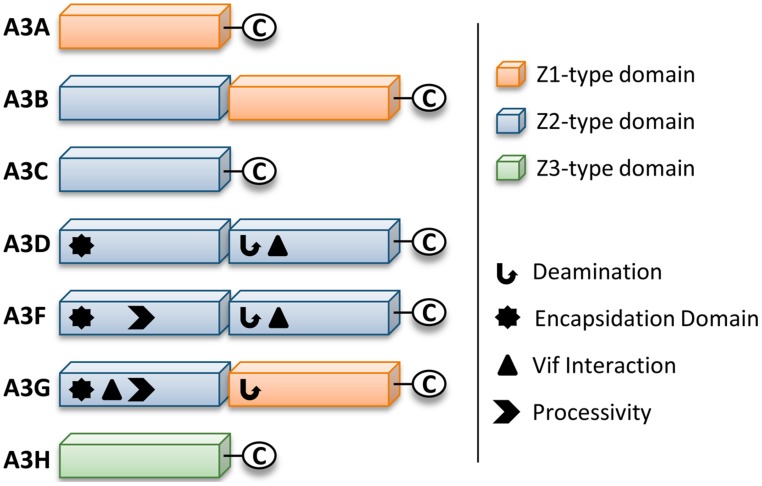
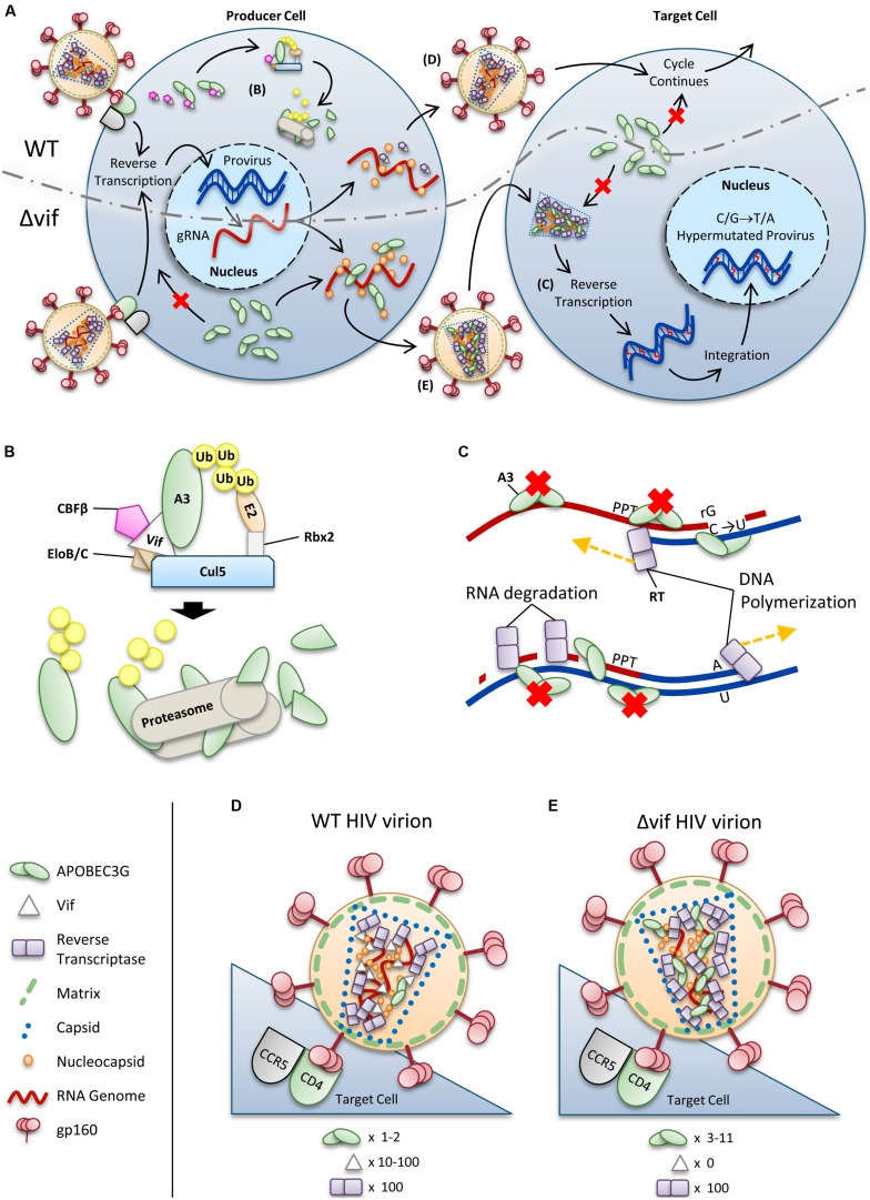
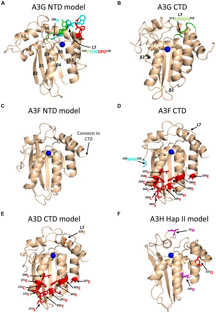
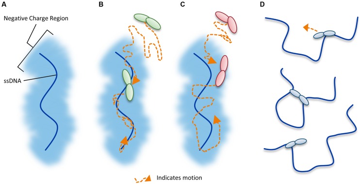
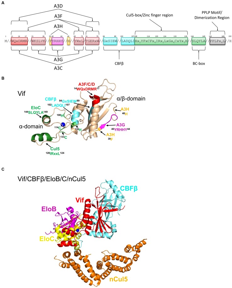
The APOBEC3 restriction factors are a family of deoxycytidine deaminases that are able to suppress replication of viruses with a single-stranded DNA intermediate by inducing mutagenesis and functional inactivation of the virus. Of the seven human APOBEC3 enzymes, only APOBEC3-D, -F, -G, and -H appear relevant to restriction of HIV-1 in CD4+ T cells and will be the focus of this review. The restriction of HIV-1 occurs most potently in the absence of HIV-1 Vif that induces polyubiquitination and degradation of APOBEC3 enzymes through the proteasome pathway. To restrict HIV-1, APOBEC3 enzymes must be encapsidated into budding virions. Upon infection of the target cell during reverse transcription of the HIV-1 RNA into (-)DNA, APOBEC3 enzymes deaminate cytosines to form uracils in single-stranded (-)DNA regions. Upon replication of the (-)DNA to (+)DNA, the HIV-1 reverse transcriptase incorporates adenines opposite to the uracils thereby inducing C/G to T/A mutations that can functionally inactivate HIV-1. APOBEC3G is the most studied APOBEC3 enzyme and it is known that Vif attempts to thwart APOBEC3 function not only by inducing its proteasomal degradation but also by several degradation-independent mechanisms, such as inhibiting APOBEC3G virion encapsidation, mRNA translation, and for those APOBEC3G molecules that still become virion encapsidated, Vif can inhibit APOBEC3G mutagenic activity. Although most Vif variants can induce efficient degradation of APOBEC3-D, -F, and -G, there appears to be differential sensitivity to Vif-mediated degradation for APOBEC3H. This review examines APOBEC3-mediated HIV restriction mechanisms, how Vif acts as a substrate receptor for a Cullin5 ubiquitin ligase complex to induce degradation of APOBEC3s, and the determinants and functional consequences of the APOBEC3 and Vif interaction from a biological and biochemical perspective.

Keywords: APOBEC3; HIV; Vif; deaminase; mutagenesis; restriction factor; ubiquitination.


Figures

FIGURE 1
FIGURE 1
Zinc (Z) coordinating-type domains of human A3 enzymes. A3 enzymes coordinate zinc through the motif H-X-E-X23-28-P-C-X2-4-C. The glutamate activates a water molecule to enable zinc-hydroxide-mediated nucleophilic attack to complete the deamination reaction. Deamination activity has been demonstrated for all A3 enzymes. For the enzymes with two Z-type domains that restrict HIV in CD4+ T cells (A3D, A3F, and A3G), a legend depicts known biochemical functions of each Z-type domain. A common feature of A3 enzymes with two Z-type domains is the segregation of functions in the N-terminal domain (NTD) and C-terminal domain (CTD). The NTD is responsible for encapsidation and the CTD is responsible for deamination activity. Both domains can bind nucleic acids. The binding site of Vif is in the NTD for A3G and in the CTD for A3D and A3F. The determinants for enzyme processivity have only been studied for A3G and A3F. A3G and A3F processivity is imparted by the NTD.
FIGURE 2
FIGURE 2
Overview of HIV restriction by A3 enzymes. (A) Sketch depicting lifecycles of wild-type (WT) and ΔVif HIV (ΔVif). Each virion enters a cell that expresses A3 enzymes. In the WT virus, Vif is expressed in the cell and recruits host cell CBFβ for stability and CRL5 E3 ubiquitin ligase complex composed of Elongin B/C (EloB/C), Cullin5 (Cul5) and Rbx2 (B). In this complex, Vif acts as the substrate receptor to induce degradation of A3 enzymes. As a result, assembling virions do not encapsidate high levels of A3 enzymes and upon infection of a target cell the HIV lifecycle continues. The ΔVif HIV encapsidates A3 enzymes through an RNA and Gag interaction. In the target cell the A3 enzymes within the capsid of HIV can deaminate cytosines to uracils in nascent single-stranded (-)DNA during reverse transcription (C). These uracils induce G→A transition mutations upon synthesis of (+)DNA (C). The resulting hypermutated virus can be integrated into the host genome but is functionally inactivated. A3 enzymes in the target cell cannot enter the HIV capsid and are unable to restrict virus replication unless encapsidated into budding virions. (B) Detailed sketch of Vif-mediated polyubiquitination of A3G. Vif interacts with Elongin C (EloC), which forms an obligate heterodimer with Elongin B (EloB), and Cul5. The transcription cofactor CBFβ stabilizes Vif. Cul5 binds to Rbx2and subsequently recruits an E2 ubiquitin conjugating enzyme. Vif is the substrate receptor that recruits A3 enzymes. The 48K-linked ubiquitin chains result in proteasomal degradation of the A3. (C) Sketch demonstrating the limited vulnerability of single-stranded (-)DNA to A3-mediated deamination that is imposed from the dynamics of reverse transcription. Reverse transcriptase is abbreviated as RT. HIV contains two polypurine tracts (PPT) that are used as primers for (+)DNA synthesis. In the figure, only one PPT is depicted. (D,E) Sketches depicting the stoichiometry of major virion components for a (D) WT and (E) ΔVif HIV virion. Figures correspond to (D) and (E) in (A). (D) Low amounts of A3 may escape Vif-mediated degradation and become virion encapsidated (approximately one to two molecules of A3G/virion). (E) A ΔVif HIV cannot induce degradation of A3 enzymes and that results in the encapsidation of A3 enzymes through an interaction with RNA and Gag. Approximately 3–11 molecules of A3G can become virion encapsidated. (D,E) Stoichiometry values for virions were obtained from Camaur and Trono (1996), Fouchier et al. (1996), Coffin et al. (1997), Xu et al. (2007), Nowarski et al. (2008).
FIGURE 3
FIGURE 3
Structures of A3 enzymes. A3 enzymes have a basic structure in each Z-type domain that is composed of a five-stranded β-sheet core surrounded by six α-helices. Numerical assignments to β-strands and α-helices are superimposed in (A). Zinc atoms are shown as blue spheres. (A) Model of the N-terminal domain (NTD) of A3G. Loop 7 (L7) of the A3G NTD is a central structure in its anti-HIV function. Highlighted on L7 are the residues important for interaction with Vif (red, 128DPD130), oligomerization/virion encapsidation (green and cyan, 124YYFW127), and jumping component of A3G processivity (cyan, 126FW127). Helix 6 (h6) is adjacent to L7 and contributes to the sliding component of A3G processivity, particularly 186H (cyan). The model of the A3G NTD was obtained by using the automated SWISS-MODEL program using the homologous A3G C-terminal domain structure (CTD, PDB: 3IQS). (B) The A3G CTD (PDB: 2KEM) is the catalytic domain of A3G. The A3G CTD has a discontinuous β2 strand forming a loop-like bulge between the β2 and β2′ strands. A3G L7 residues 313RIYDDQ318 (green) mediate tetramerization and determine the preferred deamination motif. (C) The model of the A3F NTD was obtained by using the automated SWISS-MODEL program using the homologous A3C structure (PDB: 3VM8). The end of h6 connects the NTD to the CTD and contains an 190NPM192 motif. This NPM motif is found only in A3D and A3F. (D) The A3F CTD (PDB: 4IOU) is the catalytic domain of A3F and interacts with Vif. Residues that interact with Vif across Helix 2, 3, 4, and β-strand 4 are shown in red. Also shown on this structure is the deamination motif specificity loop (L7) and the 190NPM192 motif. The structure illustrates the kinked orientation introduced by the Pro in the 190NPM192 motif, which blocks the sliding function of A3F. (E) The model of the CTD of A3D was obtained by using the SWISS-MODEL program using the homologous A3F structure (PDB: 4IOU). Residues that interact with Vif across Helix 2, 3, 4, and β-strand 4 are shown in red. The 320C residue on L7 that influences A3D activity is shown in orange. (F) Model of A3H Hap II showing residues that interact with Vif and cause haplotype instability. In A3H Hap II, 121D (red) on predicted h4 mediates an interaction with Vif. In A3H Hap I the R105G mutation induces protein instability (magenta). In A3H Hap III and IV, the deletion of 15N induces protein instability (magenta). The model of the A3H Hap II was obtained by using the automated SWISS-MODEL program using the homologous APOBEC2 structure (PDB: 2NYT). Figures were made using PyMOL (The PyMOL Molecular Graphics System, Version 1.5.05, Shrödinger, LLC.).
FIGURE 4
FIGURE 4
Illustration of DNA scanning by facilitated diffusion. (A) Sketch of DNA showing the negatively charged region of DNA important for facilitated diffusion of A3 enzymes. (B–D) Enzyme in sketches is shown as a dimer, although the oligomerization state may vary with different A3 enzymes. (B) Sketch depicting a 1-dimensional DNA scanning path by sliding. Dotted line indicates path of enzyme (orange). Sliding enables an in depth search of local areas of a substrate. (C) Sketch depicting a 3-dimensional DNA scanning path by jumping. Jumping enables larger translocations on DNA substrates, but lacks a local search process. The microdissociations of the enzyme from the DNA that occur when the enzyme jumps does not leave the negatively charged domain of the DNA so the enzyme has a higher likelihood of reassociating with the same DNA substrate than diffusion into the bulk solution. (D) Sketch depicting a 3-dimensional DNA scanning path by intersegmental transfer. Intersegmental transfer enables larger translocations on DNA substrates, but lacks a local search process. An enzyme with two DNA-binding domains binds two regions of DNA simultaneously before dissociating from one region to move to another.
FIGURE 5
FIGURE 5
Structure of Vif and host interacting partners. (A) Domain organization of Vif. Vif uses specific motifs to interact with A3G (magenta, 40YRHHY44), A3F/A3C/A3D (red, 11WQxDRMR17 and 74TGERxW79), and A3H (orange, 39F and 48H). In conjunction with these specific motifs, there are shared interaction motifs for A3F and A3G with Vif (pink, 21WKSLVK26 and 69YWxL72). CBFβ interacts with Vif through two adjacent motifs (cyan, 84GxSIEW89 and 102LADQLI107). The Zinc finger region (green, amino acids 108-139) coordinates the Zinc through an 108H114C133C139H motif and stabilizes the Vif structure, which indirectly enables an interaction with Cullin 5 (Cul5). Direct interaction of Vif with Cul5 is through amino acids 120IRxxL124. The BC box mediates an interaction with Elongin C (green 144SLQYLA149). Vif oligomerizes through a PPLP motif (gray, 163PPLPx4L169). Slanted lines are used to indicate intervening amino acids between the domains. (B) The crystal structure of Vif (PDB: 4N9F) shows that it has two domains on either side of a bound Zinc (blue). The N-terminal α/β-domain consists of a five stranded β-sheet, a discontinuous β-strand and three α-helices. The α/β-domain contains the binding interface for CBFβ (cyan, 102LADQLI107, 84GxSIEW89) and A3 enzymes. The 11WQxDRMR17 motif (red) is used to interact with A3F, A3C, and A3D, the 40YRHHY44 motif (magenta) is used to interact with A3G, and residues 39F and 48H (orange) are used to interact with A3H. The α-domain contains two alpha helices that mediate two separate interactions with EloC (green, 144SLQYLA149) and Cul5 (green, 120IRxxL124). (C) Structure HIV Vif (red) in complex with CBFβ (cyan), Elongin C (EloC, yellow), and the N-terminal domain of Cullin 5 (nCul5, amino acids 12–386, orange, PDB: 4N9F). Elongin B (EloB, magenta) dimerizes with EloC. Figures were made using PyMOL (The PyMOL Molecular Graphics System, Version 1.5.05, Shrödinger, LLC.).

References

    1. Abdel-Mohsen M., Raposo R. A., Deng X., Li M., Liegler T., Sinclair E., et al. (2013). Expression profile of host restriction factors in HIV-1 elite controllers. Retrovirology 10 106 10.1186/1742-4690-10-106 - DOI - PMC - PubMed
    1. Adolph M. B., Webb J., Chelico L. (2013). Retroviral restriction factor APOBEC3G delays the initiation of DNA synthesis by HIV-1 reverse transcriptase. PLoS ONE 8:e64196 10.1371/journal.pone.0064196 - DOI - PMC - PubMed
    1. Albin J. S., Anderson J. S., Johnson J. R., Harjes E., Matsuo H., Krogan N. J., et al. (2013). Dispersed sites of HIV Vif-dependent polyubiquitination in the DNA deaminase APOBEC3F. J. Mol. Biol. 425 1172–1182 10.1016/j.jmb.2013.01.010 - DOI - PMC - PubMed
    1. Albin J. S., Brown W. L., Harris R. S. (2014). Catalytic activity of APOBEC3F is required for efficient restriction of Vif-deficient human immunodeficiency virus. Virology 450–451, 49–54 10.1016/j.virol.2013.11.041 - DOI - PMC - PubMed
    1. Albin J. S., Hache G., Hultquist J. F., Brown W. L., Harris R. S. (2010a). Long-term restriction by APOBEC3F selects human immunodeficiency virus type 1 variants with restored Vif function. J. Virol. 84 10209–10219 10.1128/JVI.00632-10 - DOI - PMC - PubMed