. 2013 Jun 5;8(16):1491-9.
doi: 10.3969/j.issn.1673-5374.2013.16.006.

JNK3 involvement in nerve cell apoptosis and neurofunctional recovery after traumatic brain injury

Affiliations

JNK3 involvement in nerve cell apoptosis and neurofunctional recovery after traumatic brain injury

Jiang Long et al. Neural Regen Res. .

Abstract

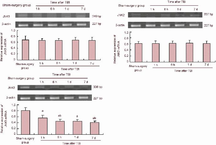
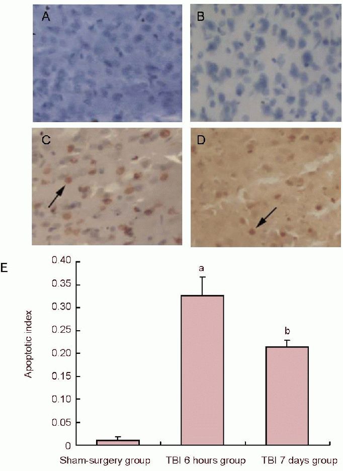
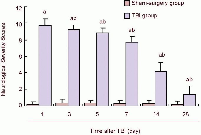
Increasing evidence has revealed that the activation of the JNK pathway participates in apoptosis of nerve cells and neurological function recovery after traumatic brain injury. However, which genes in the JNK family are activated and their role in traumatic brain injury remain unclear. Therefore, in this study, in situ end labeling, reverse transcription-PCR and neurological function assessment were adopted to investigate the alteration of JNK1, JNK2 and JNK3 gene expression in cerebral injured rats, and their role in cell apoptosis and neurological function restoration. Results showed that JNK3 expression significantly decreased at 1 and 6 hours and 1 and 7 days post injury, but that JNK1 and JNK2 expression remained unchanged. In addition, the number of apoptotic nerve cells surrounding the injured cerebral cortex gradually reduced over time post injury. The Neurological Severity Scores gradually decreased over 1, 3, 5, 14 and 28 days post injury. These findings suggested that JNK3 expression was downregulated at early stages of brain injury, which may be associated with apoptosis of nerve cells. Downregulation of JNK3 expression may promote the recovery of neurological function following traumatic brain injury.

Keywords: JNK1; JNK2; JNK3; TdT-mediated dUTP nick end labeling; cell apoptosis; neural regeneration; neurological function recovery; neuroregeneration; reverse transcription-PCR; traumatic brain injury.


Conflict of interest statement

Conflicts of interest: None declared.

Figures

Figure 1
Figure 1
Reverse transcription-PCR results of JNK1, JNK2, JNK3 expression in the peripheral area of the injured cerebral cortex after TBI in rats. aP< 0.05, vs. sham-surgery group; bP< 0.05, vs. previous time point group and 6 hours, 1 and 7 days groups. Data are expressed as mean ± SD of 10 rats from each group (one-way analysis of variance and least significant difference t-test). Sham-surgery group (gene expression expressed as the ratio of absorbance to the target gene/β-actin). TBI: Traumatic brain injury; h: hour; d: day.
Figure 2
Figure 2
Cell apoptosis in the peripheral area of the cerebral cortex in traumatic brain injury (TBI) rats (TUNEL staining). (A) Cell apoptosis of negative control (× 200); (B) morphology of apoptotic cells in the sham-surgery group (× 200); (C) cell apoptosis at 6 hours post injury (× 200); (D) morphology of apoptotic cells at 7 days post injury: the number of apoptotic cells was markedly reduced when compared to 6 hours post injury; arrows in C and D show brown positive apoptotic cells with thickening nuclei. (E) Quantification of TUNEL-positive cells. Data represent the change in cellular apoptosis index in all experimental groups (apoptosis index = number of TUNEL-positive cells/total number of cells). aP< 0.05, vs. sham-surgery group; bP< 0.05, vs. TBI 6 hours group. Data are expressed as mean ± SD of 10 rats from each group (one-way analysis of variance and least significant difference t-test). TUNEL: Terminal deoxynucleotidyl transferase-mediated dUTP-biotin nick end labeling.
Figure 3
Figure 3
Neurological Severity Scores at different time points in TBI rats. Data represent Neurological Severity Scores. Scores 13 to 18 indicate severe injury; 7 to 12, moderate injury; 1 to 6, mild injury. In TBI groups, Neurological Severe Scores gradually decreased over time, suggesting that after TBI, neurological function recovered to some extent. aP< 0.05, vs. sham-surgery group; bP< 0.05, vs. previous time points of TBI groups. Data are expressed as mean ± SD of 10 rats from each group (one-way analysis of variance and least significant difference t-test). TBI: Traumatic brain injury; d: day.

References

    1. Theadom A, Barker-Collo S, Feigin VL, et al. The spectrum captured: a methodological approach to studying incidence and outcomes of traumatic brain injury on a population level. Neuroepidemiology. 2012;38(1):18–29. - PubMed
    1. Kirov II, Tal A, Babb JS, et al. Diffuse axonal injury in mild traumatic brain injury: a 3D multivoxel proton MR spectroscopy study. J Neurol. 2013;260(1):242–252. - PMC - PubMed
    1. Dash PK, Kobori N, Moore AN. A molecular description of brain trauma pathophysiology using microarray technology: an overview. Neurochem Res. 2004;29(6):1275–1286. - PubMed
    1. Brown R, Thompson HJ, Imran SA, et al. Traumatic brain injury induces adipokine gene expression in rat brain. Neurosci Lett. 2008;432(1):73–78. - PMC - PubMed
    1. Raghupathi R. Cell death mechanisms following traumatic brain injury. Brain Pathol. 2004;14(2):215–222. - PMC - PubMed