Review
. 2013 Jul 25;8(21):2003-14.
doi: 10.3969/j.issn.1673-5374.2013.21.009.

Oxidative stress, mitochondrial damage and neurodegenerative diseases

Affiliations
Review

Oxidative stress, mitochondrial damage and neurodegenerative diseases

Chunyan Guo et al. Neural Regen Res. .

Abstract

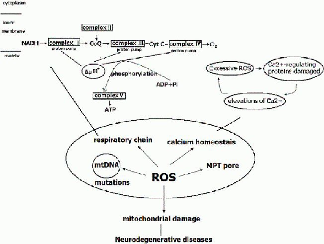
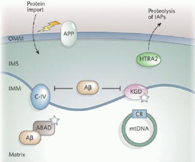
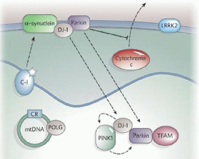
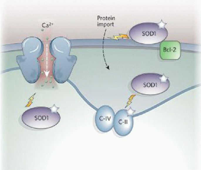
Oxidative stress and mitochondrial damage have been implicated in the pathogenesis of several neurodegenerative diseases, including Alzheimer's disease, Parkinson's disease and amyotrophic lateral sclerosis. Oxidative stress is characterized by the overproduction of reactive oxygen species, which can induce mitochondrial DNA mutations, damage the mitochondrial respiratory chain, alter membrane permeability, and influence Ca(2+) homeostasis and mitochondrial defense systems. All these changes are implicated in the development of these neurodegenerative diseases, mediating or amplifying neuronal dysfunction and triggering neurodegeneration. This paper summarizes the contribution of oxidative stress and mitochondrial damage to the onset of neurodegenerative eases and discusses strategies to modify mitochondrial dysfunction that may be attractive therapeutic interventions for the treatment of various neurodegenerative diseases.

Keywords: Alzheimer's disease; Parkinson's disease; amyotrophic lateral sclerosis; grants-supported paper; mitochondrial damage; neural regeneration; neurodegenerative diseases; neuroregeneration; oxidative stress; reactive oxygen species; respiratory chain.


Conflict of interest statement

Conflict of Interest: None declared.

Figures

Figure 1
Figure 1
Reactive oxygen species (ROS) and mitochondrial damage. The mitochondrial respiratory chain complexes consist of five complexes: complexes I, II, III, IV and V, which catalyze the phosphorylation of adenosine diphosphate (ADP) to adenosine triphosphate (ATP). Complex I: nicotinamide adenine dinucleotide (NADH)-coenzyme Q; Complex II: succinate dehydrogenase-coenzyme Q; Complex III: coenzyme Q-cytochrome c reductase; Complex IV: cytochrome c oxidase; Complex V: ATP synthase. More than 80 proteins comprise these complexes, 13 of them encoded by mitochondrial DNA. Complex I, II, III, and IV constitute the electron-transport chain. The electrons, borne on NAD+, are transferred to NADH and then to coenzyme Q, and the electrons are transferred to complex II and to coenzyme Q. From coenzyme Q, the electrons are passed to complex III, then to cytochrome c, then to complex IV and finally to 1/2 O2 to give H2O. ATP is generated by the influx of these protons back into the mitochondrial matrix through ATP synthase. The highly ROS can damage NADH dehydrogenase, cytochrome c oxidase, and ATP synthase, resulting in shutdown of mitochondrial energy production. Under normal physiological conditions, Ca2+ fluxes are properly controlled across the plasma membrane and between intracellular compartments. Excessive ROS generation can directly damage Ca2+-regulating proteins (in the plasma membrane, such as ligand- and voltage-gated Ca2+ channels, endoplasmic reticulum Ca2+-ATP synthases, and mitochondria electron-transport chain proteins), resulting in elevations of Ca2+, which disturb Ca2+ homeostasis. Overproduction of ROS can lead to mitochondrial damage, including mutations in mitochondrial DNA, damage to the mitochondrial respiratory chain and mitochondrial membrane permeability, and disruption to Ca2+ homeostasis. Therefore, mitochondrial damage plays an important role in the pathogenesis of neurodegenerative diseases. CoQ: Coenzyme Q; Cyt c: cytochrome c; mtDNA: mitochondrial DNA; MPT: mitochondrial permeability transition.
Figure 2
Figure 2
The role of mitochondria in Alzheimer's disease[1]. Mitochondrial reactive oxygen species (ROS) generation can increase β-amyloid protein (Aβ) levels, and Aβ can interact with mitochondria and further cause mitochondrial dysfunction. Aβ inhibits mitochondrial respiratory chain complex IV (C-IV) and α-ketoglutarate dehydrogenase (KGD), and binds Aβ-binding alcohol dehydrogenase (ABAD). Both KGD and ABAD produce ROS (white stars). The amyloid precursor protein (APP) may be targeted to the outer mitochondrial membrane (OMM) and interfere with protein import. Mitochondria have also been reported to contain active γ-secretase complexes, which are involved in cleaving the APP to form Aβ and contain presenilin 1, which increases the proteolytic activity of high temperature requirement protein A2 (HTRA2) towards the inhibitor of apoptosis proteins (IAPs). Alzheimer's disease patients have on average more somatic mutations in the mitochondrial DNA (mtDNA) control region than control subjects. IMM: Inner mitochondrial membrane; IMS: intermembrane space.
Figure 3
Figure 3
The role of mitochondria in Parkinson's disease[1]. Complex I (C-I) activity is decreased in Parkinson's disease, and inhibition of C-I by mitochondrial permeability transition pore or rotenone causes Parkinsonism. Mutations in mitochondrial DNA (mtDNA)-encoded C-I subunits and mtDNA polymerase γ (POLG) also cause Parkinsonism. Many genes associated with Parkinson's disease (such as α-synuclein, parkin, DJ-1, PINK1, and LRRK2) also implicate mitochondria in disease pathogenesis. α-synuclein overexpression can impair mitochondrial function and enhance the toxicity of the mitochondrial permeability transition pore. Parkin associates with the outer mitochondrial membrane and protects against cytochrome c release. It may also associate with mitochondrial-transcription-factor A (TFAM). Physical associations have been reported between DJ-1 and α-synuclein, DJ-1 and PINK1, and DJ-1 and parkin, and there is genetic evidence that DJ-1, PINK1 and parkin are functionally linked in the same pathway. About 10% of the kinase LRRK2 is localized to mitochondria, and Parkinson's disease-related mutations augment its kinase activity. A mutation in high temperature requirement protein A2 was found in 1% of sporadic Parkinson's disease patients. CR: Control region.
Figure 4
Figure 4
The role of mitochondria in amyotrophic lateral sclerosis[1]. Overexpression of mutant superoxide dismutase 1 (SOD1) in amyotrophic lateral sclerosis impairs electron transport chain activities and decreases mitochondrial calcium-loading capacity. SOD1 has been localized to the outer mitochondrial membrane, intermembrane space and matrix, and targeting of mutant SOD1 to mitochondria causes cytochrome c release and apoptosis. Mutant SOD1 promotes aberrant mitochondrial reactive oxygen species production and forms aggregates that may clog the outer mitochondrial membrane protein importation machinery or bind and sequester the antiapoptotic protein Bcl-2. C-II: Complex II; C-IV: complex IV.

References

    1. Lin MT, Beal MF. Mitochondrial dysfunction and oxidative stress in neurodegenerative diseases. Nature. 2006;443(7113):787–795. - PubMed
    1. Martin LJ. Mitochondrial and cell death mechanisms in neurodegenerative diseases. Pharmaceuticals (Basel) 2010;3(4):839–915. - PMC - PubMed
    1. Crunkhorn S. Neurodegenerative disorders: Restoring the balance. Nat Rev Drug Discov. 2011;10(8):576. - PubMed
    1. Sas K, Robotka H, Toldi J, et al. Mitochondria, metabolic disturbances, oxidative stress and the kynurenine system, with focus on neurodegenerative disorders. J Neurol Sci. 2007;257(1-2):221–239. - PubMed
    1. Douarre C, Sourbier C, Dalla Rosa I, et al. Mitochondrial topoisomerase I is critical for mitochondrial integrity and cellular energy metabolism. PLoS One. 2012;7(7):e41094. - PMC - PubMed