

Buyang Huanwu Decoction regulates neural stem cell behavior in ischemic brain
- PMID: 25206543
- PMCID: PMC4146048
- DOI: 10.3969/j.issn.1673-5374.2013.25.004
Buyang Huanwu Decoction regulates neural stem cell behavior in ischemic brain
Abstract
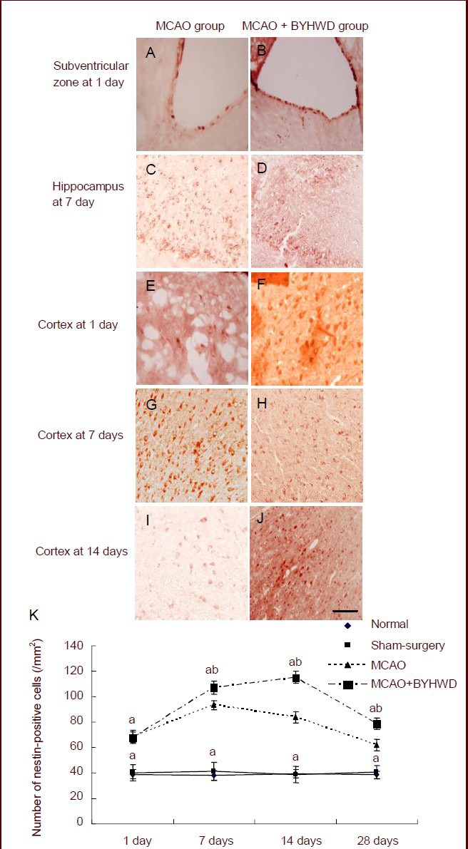
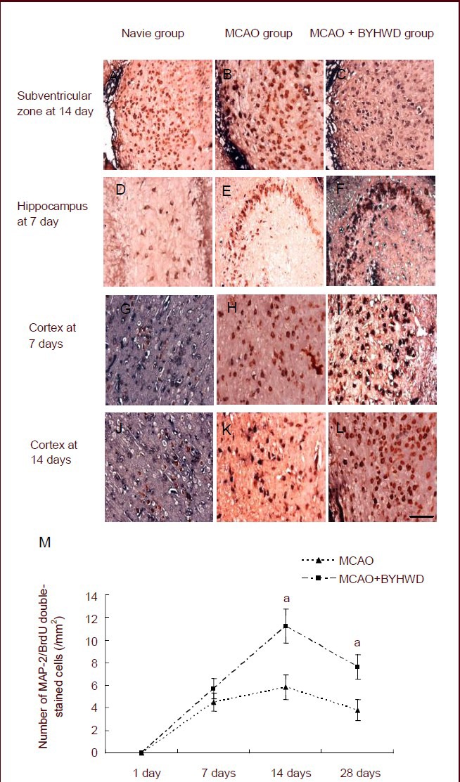
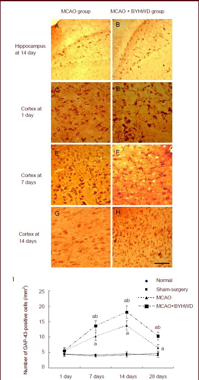
The traditional Chinese medicine Buyang Huanwu Decoction has been shown to improve the neu-rological function of patients with stroke. However, the precise mechanisms underlying its effect remain poorly understood. In this study, we established a rat model of cerebral ischemia by middle cerebral artery occlusion and intragastrically administered 5 g/kg Buyang Huanwu Decoction, once per day, for 1, 7, 14 and 28 days after cerebral ischemia. Immunohistochemical staining revealed a number of cells positive for the neural stem cell marker nestin in the cerebral cortex, the subven-tricular zone and the ipsilateral hippocampal dentate gyrus in rat models of cerebral ischemia. Buyang Huanwu Decoction significantly increased the number of cells positive for 5-bromodeoxyuridine (BrdU), a cell proliferation-related marker, microtubule-associated protein-2, a marker of neuronal differentiation, and growth-associated protein 43, a marker of synaptic plasticity in the ischemic rat cerebral regions. The number of positive cells peaked at 14 and 28 days after intragastric administration of Buyang Huanwu Decoction. These findings suggest that Buyang Huanwu Decoction can promote the proliferation and differentiation of neural stem cells and hance synaptic plasticity in ischemic rat brain tissue.
Keywords: BrdU; Buyang Huanwu Decoction; cerebral cortex; cerebral ischemia; dentate gyrus; differentiation; grants-supported paper; growth-associated protein 43; microtubule-associated protein-2; nestin; neural regeneration; neural stem cells; neuroregeneration; proliferation; subventricular zone; traditional Chinese medicine.
Conflict of interest statement
Figures
Similar articles
-
Buyang Huanwu decoction facilitates neurorehabilitation through an improvement of synaptic plasticity in cerebral ischemic rats.BMC Complement Altern Med. 2017 Mar 28;17(1):173. doi: 10.1186/s12906-017-1680-9. BMC Complement Altern Med. 2017. PMID: 28351388 Free PMC article.
-
[Buyang Huanwu Decoction attenuates cerebral ischemia-reperfusion injury in rats via miR-26a-5p mediated PTEN/PI3K/Akt signaling pathway].Zhongguo Zhong Yao Za Zhi. 2024 Aug;49(15):4197-4206. doi: 10.19540/j.cnki.cjcmm.20240515.501. Zhongguo Zhong Yao Za Zhi. 2024. PMID: 39307758 Chinese.
-
Buyang Huanwu Decoction fraction protects against cerebral ischemia/reperfusion injury by attenuating the inflammatory response and cellular apoptosis.Neural Regen Res. 2013 Jan 25;8(3):197-207. doi: 10.3969/j.issn.1673-5374.2013.03.001. Neural Regen Res. 2013. PMID: 25206589 Free PMC article.
-
[Effects of Buyang Huanwu Decoction on neurovascular units after cerebral ischemia: a review].Zhongguo Zhong Yao Za Zhi. 2021 Oct;46(20):5226-5232. doi: 10.19540/j.cnki.cjcmm.20210610.706. Zhongguo Zhong Yao Za Zhi. 2021. PMID: 34738423 Review. Chinese.
-
A Review of Traditional Chinese Medicine, Buyang Huanwu Decoction for the Treatment of Cerebral Small Vessel Disease.Front Neurosci. 2022 Jun 29;16:942188. doi: 10.3389/fnins.2022.942188. eCollection 2022. Front Neurosci. 2022. PMID: 35844225 Free PMC article. Review.
Cited by
-
Therapeutic Effect of Buyang Huanwu Decoction on the Gut Microbiota and Hippocampal Metabolism in a Rat Model of Cerebral Ischemia.Front Cell Infect Microbiol. 2022 Jun 14;12:873096. doi: 10.3389/fcimb.2022.873096. eCollection 2022. Front Cell Infect Microbiol. 2022. PMID: 35774407 Free PMC article.
-
The Effect of Traditional Chinese Medicine on Neural Stem Cell Proliferation and Differentiation.Aging Dis. 2017 Dec 1;8(6):792-811. doi: 10.14336/AD.2017.0428. eCollection 2017 Dec. Aging Dis. 2017. PMID: 29344417 Free PMC article. Review.
-
What can traditional Chinese medicine do for adult neurogenesis?Front Neurosci. 2023 Apr 12;17:1158228. doi: 10.3389/fnins.2023.1158228. eCollection 2023. Front Neurosci. 2023. PMID: 37123359 Free PMC article. Review.
-
Buyang Huanwu Decoction () Attenuates Glial Scar by Downregulating the Expression of Leukemia Inhibitory Factor in Intracerebral Hemorrhagic Rats.Chin J Integr Med. 2019 Apr;25(4):264-269. doi: 10.1007/s11655-018-2917-7. Epub 2018 Dec 28. Chin J Integr Med. 2019. PMID: 30607786
-
Efficacy and safety of Buyang Huanwu Decoction in the treatment of post-stroke depression: A systematic review and meta-analysis of 15 randomized controlled trials.Front Neurol. 2022 Nov 4;13:981476. doi: 10.3389/fneur.2022.981476. eCollection 2022. Front Neurol. 2022. PMID: 36408491 Free PMC article.
References
-
- Curtis MA, Kam M, Nannmark U, et al. Human neuroblasts migrate to the olfactory bulb via a lateral ventricular extension. Science. 2007;315(5816):1243–1249. - PubMed
-
- Thored P, Arvidsson A, Cacci E, et al. Persistent production of neurons from adult brain stem cells during recovery after stroke. Stem Cells. 2006;24(3):739–747. - PubMed
-
- Ormerod BK, Galea LA. Reproductive status influences cell proliferationand cell survival in the dentate gyrus of adult female meadowvoles: a possible regulatory role for estradiol. Neuroscience. 2001;102(2):369–379. - PubMed
LinkOut - more resources
Full Text Sources
Research Materials
Miscellaneous