3D ultrafast ultrasound imaging in vivo
- PMID: 25207828
- PMCID: PMC4820600
- DOI: 10.1088/0031-9155/59/19/L1
3D ultrafast ultrasound imaging in vivo
Abstract
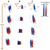
Very high frame rate ultrasound imaging has recently allowed for the extension of the applications of echography to new fields of study such as the functional imaging of the brain, cardiac electrophysiology, and the quantitative imaging of the intrinsic mechanical properties of tumors, to name a few, non-invasively and in real time. In this study, we present the first implementation of Ultrafast Ultrasound Imaging in 3D based on the use of either diverging or plane waves emanating from a sparse virtual array located behind the probe. It achieves high contrast and resolution while maintaining imaging rates of thousands of volumes per second. A customized portable ultrasound system was developed to sample 1024 independent channels and to drive a 32 × 32 matrix-array probe. Its ability to track in 3D transient phenomena occurring in the millisecond range within a single ultrafast acquisition was demonstrated for 3D Shear-Wave Imaging, 3D Ultrafast Doppler Imaging, and, finally, 3D Ultrafast combined Tissue and Flow Doppler Imaging. The propagation of shear waves was tracked in a phantom and used to characterize its stiffness. 3D Ultrafast Doppler was used to obtain 3D maps of Pulsed Doppler, Color Doppler, and Power Doppler quantities in a single acquisition and revealed, at thousands of volumes per second, the complex 3D flow patterns occurring in the ventricles of the human heart during an entire cardiac cycle, as well as the 3D in vivo interaction of blood flow and wall motion during the pulse wave in the carotid at the bifurcation. This study demonstrates the potential of 3D Ultrafast Ultrasound Imaging for the 3D mapping of stiffness, tissue motion, and flow in humans in vivo and promises new clinical applications of ultrasound with reduced intra--and inter-observer variability.
Figures


References
-
- Bercoff J, Tanter M, Fink M. Supersonic shear imaging: a new technique for soft tissue elasticity mapping. IEEE Trans. Ultrason. Ferroelectr. Freq. Control. 2004;51:396–409. doi:10.1109/TUFFC.2004.1295425. - PubMed
-
- Couade M, Pernot M, Prada C, Messas E, Emmerich J, Bruneval P, Criton A, Fink M, Tanter M. Quantitative Assessment of Arterial Wall Biomechanical Properties Using Shear Wave Imaging. Ultrasound Med. Biol. 2010;36:1662–1676. doi:10.1016/j.ultrasmedbio.2010.07.004. - PubMed
-
- Couade M, Pernot M, Tanter M, Messas E, Bel A, Ba M, Hagege A-A, Fink M. Ultrafast imaging of the heart using circular wave synthetic imaging with phased arrays. Ultrasonics Symposium (IUS), 2009 IEEE International; Presented at the Ultrasonics Symposium (IUS), 2009 IEEE International; 2009; pp. 515–518. doi:10.1109/ULTSYM.2009.5441640.
-
- Denarie B, Bjastad T, Torp H. Multi-line transmission in 3-D with reduced crosstalk artifacts: a proof of concept study. IEEE Trans. Ultrason. Ferroelectr. Freq. Control. 2013;60:1708–1718. doi:10.1109/TUFFC.2013.2752. - PubMed
Publication types
MeSH terms
Grants and funding
LinkOut - more resources
Full Text Sources
Other Literature Sources
