. 2014 Jul-Aug;20(4):403-12.
doi: 10.15274/INR-2014-10041. Epub 2014 Aug 28.

Selective and superselective angiography of pediatric moyamoya disease angioarchitecture in the posterior circulation

Affiliations

Selective and superselective angiography of pediatric moyamoya disease angioarchitecture in the posterior circulation

Gerasimos Baltsavias et al. Interv Neuroradiol. 2014 Jul-Aug.

Abstract

The anastomotic network of the posterior circulation in children with moyamoya disease has not been analyzed. We aimed to investigate the angiographic anatomy of this unique vascular network in patients with childhood moyamoya disease. Selective and superselective injections of the posterior circulation were performed in six children with newly diagnosed moyamoya disease. The arterial branches feeding the moyamoya anastomotic network, their connections and the recipient vessels were demonstrated. Depending on the level of the steno-occlusive lesion, the feeding vessels were the thalamoperforators, the posterior choroidals, the splenic artery, parietoccipital artery, other cortical posterior cerebral artery (PCA) branches, the dural branch of the PCA, the premamillary artery and other posterior communicating artery perforators. Through connections, which are described, the recipient vessels were the striate and medullary arteries, other thalamic arteries with or without medullary extensions, the pericallosal artery, medial parietoccipital cortical branches of the PCA and the anterior choroidal artery. High quality selective and superselective angiography helped in demonstrating the angiographic anatomy of the moyamoya posterior anastomotic network previously either vaguely or incompletely described, as well as connections within the posterior circulation but also its relevance as a collateral to the anterior circulation.

Keywords: digital subtraction angiography; moyamoya collateral networks; moyamoya vessels; pediatric moyamoya disease.


Figures

Figure 1
Figure 1
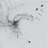
Anteroposterior (A) and lateral (B) views of a superselective injection of a thalamoperforator-peduncular perforator (short thin arrow) common ostium. The thalamoperforator (small arrowheads) courses anteriorly and laterally and at the level of the thalamocaudate sulcus anastomoses with striatal vessels which opacify an intrastriatal network (thin long arrows). Then a proximal striate artery (thick long arrow) is opacified retrogradely as well as the medullary system at the level of the angle of the lateral ventricle (thick arrowheads).
Figure 2
Figure 2
AP (A) and lateral (B) views of a superselective thalamoperforator injection (arrow) showing an extensive collateralization to distal MCA cortical branches (large arrowheads) with retrograde flow through medullary arteries (thin arrows) connected with the dilated thalamoperforator at the level of the lateral ventricular wall (small arrowheads).
Figure 3
Figure 3
A) Anteroposterior view of a vertebral injection showing an intrathalamic network (long thin arrows) of dilated vessels supplied among other branches, by a left-sided thalamoperforator (arrowheads) in a patient with occlusion of the ipsilateral PCA beyond the P2 segment. B) The same injection in lateral projection showing the thalamoperforator (short arrows) connected with the intrathalamic network. The pericallosal artery (large arrowhead) is reconstructed through several septal transcallosal collaterals (small arrowheads) projecting above the level of the thalamic network and most likely supplied by the midline choroidal arteries.
Figure 4
Figure 4
Lateral view of a superselective injection of the left P2 segment of another patient (large arrowhead at the basilar tip). The choroidal arteries (small arrowheads) at the level of the foramen of Monroe clearly the supply septal and transcallosal arterial branches (long arrows) which reconstruct the pericallosal artery (short arrows).
Figure 5
Figure 5
Lateral view of a vertebral injection showing the posterior choroidals (arrowheads) supplying both midline transcallosal branches (short arrows) which reconstruct the pericallosal artery (short thick arrows), as well as lateral branches along the floor of the lateral ventricle (thin long arrows) supplying distal striatal branches and retrogradely a single lateral striatal artery (long thick arrow).
Figure 6
Figure 6
Anteroposterior (A) and lateral (B) views of a superselective injection of the left Pcom (thick arrowhead) which through Pcom perforators (small arrowheads) opacifies the anterior choroidal artery (small short arrows) and reconstructs the MCA (long thick arrows) via the anastomotic connection of the uncal artery (long thin arrows).
Figure 7
Figure 7
Anteroposterior (A) and lateral (B) views of a right ICA injection. The distal ICA (large arrowhead) is highly stenotic. The anterior choroidal is still patent with anterograde flow and the Pcom (long thick arrow) mainly supplies a dilated tuberothalamic artery (long thin arrows) which runs superiorly along the lateral wall of the posterior hypothalamus and at the level of the terminal sulcus turns lateral on the ventricular surface of the caudate nucleus where it anastomoses with the distal segment of a lateral striate artery (small arrowheads). Further opacification of the medullary system (short arrows) is better visible in the later phases (C,D).
Figure 8
Figure 8
Anteroposterior (A) and lateral (B) views of a superselective injection of a proximal Pcom hypothalamic perforator (long thick arrow) showing collateral supply to both ACA (short thin arrows) and MCA territories. The ACA is supplied at the level of Acom with retrograde flow through hypothalamic perforators connected with the injected perforator at the level of the 3rd ventricle wall (antero-inferior arrowheads). Instead, the MCA territory is supplied with retrograde flow through the medullary arteries (long thin arrows), at the level of the angle of the lateral ventricle (thick arrowhead), fed by the distal branches of a striate artery (short thick arrows), connected (posterosuperior arrowheads) with the injected perforator.
Figure 9
Figure 9
Lateral (A) and anteroposterior (B) views of a superselective PCA injection and its dural branch (arrowheads) which supplies another dural branch (long thin arrows) roughly parallel to the straight sinus and then through fine leptomeningeal vessels (short arrows) reconstructs the superficial medial occipital cortical arteries bilaterally. Small short arrow at the basilar tip.

References

    1. Mugikura S, Takahashi S, Higano S, et al. Predominant involvement of ipsilateral anterior and posterior circulations in moyamoya disease. Stroke. 2002;33(6):1497–1500. doi: 10.1161/01.STR.0000016828.62708.21. - DOI - PubMed
    1. Mugikura S, Higano S, Shirane R, et al. Posterior circulation and high prevalence of ischemic stroke among young pediatric patients with Moyamoya disease: evidence of angiography-based differences by age at diagnosis. Am J Neuroradiol. 2011;32(1):192–198. doi: 10.3174/ajnr.A2216. - DOI - PMC - PubMed
    1. Satoh S, Shibuya H, Matsushima Y, et al. Analysis of the angiographic findings in cases of childhood moyamoya disease. Neuroradiology. 1988;30(2):111–119. doi: 10.1007/BF00395611. - DOI - PubMed
    1. Suzuki J, Takaku A. Cerebrovascular "moyamoya" disease. Disease showing abnormal net-like vessels in base of brain. Arch Neurol. 1969;20(3):288–299. doi: 10.1001/archneur.1969.00480090076012. - DOI - PubMed
    1. Hasuo K, Tamura S, Kudo S, et al. Moya moya disease: use of digital subtraction angiography in its diagnosis. Radiology. 1985;157(1):107–111. - PubMed

LinkOut - more resources