

Longitudinal changes in peripapillary atrophy in the ocular hypertension treatment study: a case-control assessment
- PMID: 25208858
- PMCID: PMC4682350
- DOI: 10.1016/j.ophtha.2014.07.033
Longitudinal changes in peripapillary atrophy in the ocular hypertension treatment study: a case-control assessment
Abstract
Purpose: To explore the association between peripapillary atrophy (PPA) area and conversion from ocular hypertension (OHT) to glaucoma.
Design: Prospective, longitudinal cohort study of cases and controls.
Participants: We included 279 age-matched and follow-up time-matched eyes with OHT that converted to glaucoma and 279 eyes with OHT that did not convert to glaucoma.
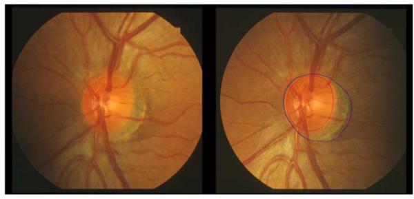
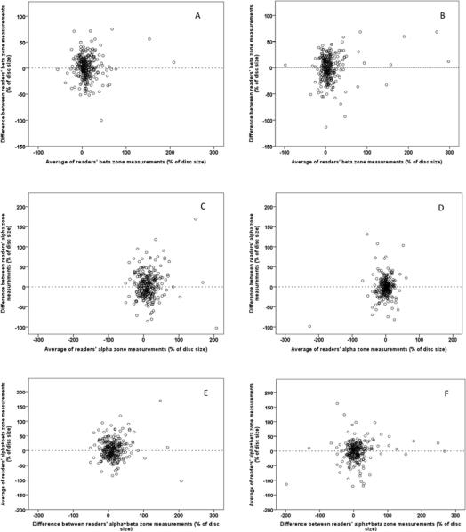
Methods: Initial and last acceptable optic disc photos were analyzed. Disc, α-zone, and β-zone PPA were traced independently by 2 trained readers and their areas were measured with Photoshop. The α-zone and β-zone areas were expressed as a percentage of optic disc area.
Main outcome measures: α-Zone and β-zone PPA size over time.
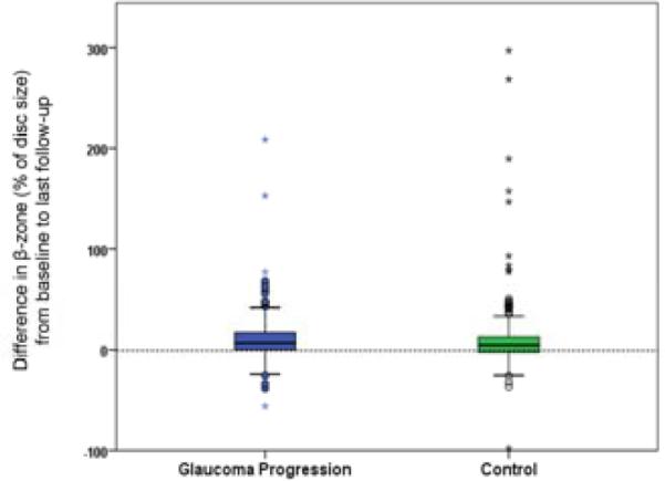
Results: Intraclass correlation coefficients (ICCs) demonstrated that readers had good agreement on disc area (ICC = 0.97) and β-zone (ICC = 0.82), but not α-zone (ICC = 0.48). The ß-zone, as a percentage of disc area, increased in size (P < 0.001) in both eyes with incident primary open-angle glaucoma (mean, 10.6%; standard deviation, 22.6%) and matched controls (mean, 10.1%; standard deviation, 33.7) over follow-up (mean, 12.3 years). The increase in size did not differ between cases and controls (P = 0.82). Enlargement of the β-zone was not correlated with follow-up time (P = 0.39).
Conclusions: The results did not show a difference in size of the β-zone at baseline between eyes that proceed to develop glaucoma and those that do not. Moreover, the β-zone enlarges equally in case and control eyes during follow-up.
Copyright © 2015 American Academy of Ophthalmology. Published by Elsevier Inc. All rights reserved.
Figures
References
-
- Fingeret M, Medeiros FA, Susanna R, Jr, Weinreb RN. Five rules to evaluate the optic disc and retinal nerve fiber layer for glaucoma. Optometry. 2005;76:661–8. - PubMed
-
- Susanna R, Jr, Vessani RM. New findings in the evaluation of the optic disc in glaucoma diagnosis. Curr Opin Ophthalmol. 2007;18:122–8. - PubMed
-
- Derick RJ, Pasquale LR, Pease ME, Quigley HA. A clinical study of peripapillary crescents of the optic disc in chronic experimental glaucoma in monkey eyes. Arch Ophthalmol. 1994;112:846–50. - PubMed
-
- See JL, Nicolela MT, Chauhan BC. Rates of neuroretinal rim and peripapillary atrophy area change: a comparative study of glaucoma patients and normal controls. Ophthalmology. 2009;116:840–7. - PubMed
-
- Quigley HA, Katz J, Derick RJ, et al. An evaluation of optic disc and nerve fiber layer examinations in monitoring progression of early glaucoma damage. Ophthalmology. 1992;99:19–28. - PubMed
Publication types
MeSH terms
Grants and funding
LinkOut - more resources
Full Text Sources
Other Literature Sources
Medical
