. 2014 Oct;141(19):3709-20.
doi: 10.1242/dev.112185. Epub 2014 Sep 10.

Semaphorin 6B acts as a receptor in post-crossing commissural axon guidance

Affiliations

Semaphorin 6B acts as a receptor in post-crossing commissural axon guidance

Irwin Andermatt et al. Development. 2014 Oct.

Abstract

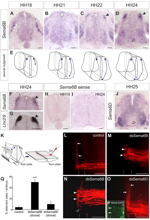
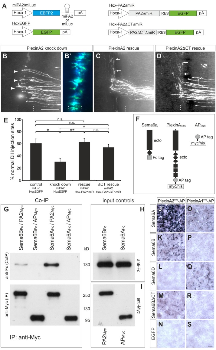
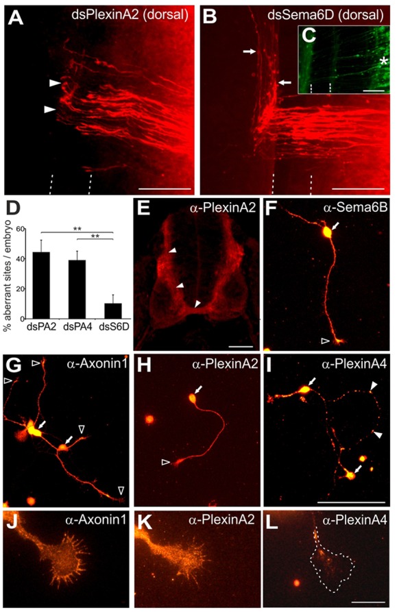
Semaphorins are a large family of axon guidance molecules that are known primarily as ligands for plexins and neuropilins. Although class-6 semaphorins are transmembrane proteins, they have been implicated as ligands in different aspects of neural development, including neural crest cell migration, axon guidance and cerebellar development. However, the specific spatial and temporal expression of semaphorin 6B (Sema6B) in chick commissural neurons suggested a receptor role in axon guidance at the spinal cord midline. Indeed, in the absence of Sema6B, post-crossing commissural axons lacked an instructive signal directing them rostrally along the contralateral floorplate border, resulting in stalling at the exit site or even caudal turns. Truncated Sema6B lacking the intracellular domain was unable to rescue the loss-of-function phenotype, confirming a receptor function of Sema6B. In support of this, we demonstrate that Sema6B binds to floorplate-derived plexin A2 (PlxnA2) for navigation at the midline, whereas a cis-interaction between PlxnA2 and Sema6B on pre-crossing commissural axons may regulate the responsiveness of axons to floorplate-derived cues.

Keywords: Chick; Cis-interaction; In ovo RNAi; PlexinA2; Spinal cord development.


Figures

Fig. 1.
Fig. 1.
Sema6b expression in dI1 commissural neurons peaks at HH24 and is required for post-crossing commissural axon guidance. (A-E) Expression of Sema6b mRNA in transverse sections of the chicken lumbar spinal cord at the indicated developmental stages (A-D) with schematic representations of the corresponding growth of dI1 axons (E). Sema6b is first detected in dI1 neurons at HH22, when their axons have reached the ipsilateral floorplate border (C, arrowhead). Expression of Sema6b in dI1 neurons is strongest at HH24 (D, arrowheads). (F,G) Probing adjacent sections with Lhx2/9 confirmed that Sema6b is expressed in dI1 neurons (arrowheads). (H,I) No staining is seen with a Sema6b sense probe. (J) Diffuse expression of Sema6d at HH25, with higher levels in motoneurons. Neither Sema6b nor Sema6d is expressed in the floorplate (D,J, open arrowheads). (K) Schematic of an open-book preparation and application of DiI for axonal tracing (red). D, dorsal; V, ventral; R, rostral; C, caudal. (L) Commissural axons cross the midline and extend along the contralateral floorplate border (arrows) in untreated control embryos. (M,N) After silencing Sema6B by injection and electroporation of dsRNA (dsSema6b), axons stall at the contralateral floorplate border (closed arrowheads) or even turn caudally (open arrowhead). (O) Silencing Sema6D by injection and electroporation of dsRNA (dsSema6d) did not affect axon guidance (arrows). (P) Expression of a co-electroporated EGFP reporter confirmed the exclusively dorsal targeting of dsRNA (asterisks). Arrows mark axons of contralaterally projecting commissural neurons. The floorplate is indicated by dashed lines. (Q) Quantification of DiI injection sites with aberrant axonal pathfinding. ***P<0.001; error bars indicate s.e.m. Scale bars: 50 µm in A-D,F-J; 100 µm in L-P.
Fig. 2.
Fig. 2.
The intracellular domain of Sema6B is essential for post-crossing commissural axon guidance. (A) Schematics of miRNA and rescue constructs used in B-H. (B-G″) Open-book analysis of DiI injection sites in embryos co-electroporated with: (B) miLuc and pMES; (C) miS6B and pMES; (D) miS6B and pSema6BΔmiR; (E) miS6B and pSema6BΔCTΔmiR; (F) pSema6BΔmiR alone; or (G) pSema6BΔCTΔmiR alone. Arrows (B,D,F) indicate normal crossing and turning of commissural axons. Post-crossing axons that failed to turn correctly at the contralateral floorplate border are indicated by arrowheads (C,E,G). (B′-G′) Merge of DiI-labeled axons (red) and EGFP (green) used to visualize the expression of pMES constructs. (B″-G″) Enhanced blue fluorescent protein-2 (EBFP2) visualizes the expression of miRNA constructs. The floorplate is indicated by dashed lines. Scale bars: 100 µm. (H) Quantification of DiI injection sites with normal axonal pathfinding. **P<0.01, ***P<0.001; n.s., not significant; error bars indicate s.e.m.
Fig. 3.
Fig. 3.
For correct pathfinding, PlxnA2 is required in both commissural neurons and the floorplate. (A-D′) Expression patterns of plexins analyzed by in situ hybridization at stages HH21 (A-D) and HH25 (A′-D′). Staining in commissural neurons is indicated by closed arrowheads. Motoneurons are marked with asterisks. The floorplate is indicated by open arrowheads and shown at higher magnification in the insets. (E-G) After downregulation of PlxnA1 (E), PlxnA2 (F) or PlxnA4 (G) by unilateral electroporation of dsRNA, axons failed to turn rostrally at the contralateral floorplate border (arrowheads). (H) Only injection sites in the electroporated area (verified by EGFP expression) were included in the analysis. (I-K) Analysis of commissural axon pathfinding after downregulation of PlxnA2 (I), PlxnA4 (J) or PlxnC1 (K) exclusively in the ventral spinal cord. After ventral downregulation of PlxnA2 (I, arrowheads), post-crossing axons failed to turn into the longitudinal axis or even turned caudally. Axon guidance was unaffected after ventral downregulation of PlxnA4 (J, arrow) or PlxnC1 (K, arrow). (L) Successful and exclusive targeting of cells at the ventral midline was verified by EGFP expression. The floorplate is indicated by dashed lines. (M,N) Quantification of injection sites with aberrant axonal pathfinding after (M) unilateral or (N) ventral electroporation of dsRNA. ***P<0.001; **P<0.01; error bars indicate s.e.m. Scale bars: 50 μm in A-D′; 100 µm in E-L.
Fig. 4.
Fig. 4.
The extracellular domain of PlxnA2 mediates its axon guidance activity in the floorplate and binds to Sema6B. (A) Schematics of the constructs used in B-E. Hoxa1 drives floorplate-specific expression. (B-D) Open-book analysis of embryos co-electroporated with: (B) miPA2 and Hox-EGFP; (C) miPA2 and Hox-PA2ΔmiR; or (D) miPA2 and Hox-PA2ΔCTΔmiR. (B′) The successful and exclusive targeting of floorplate cells was confirmed by EGFP and EBFP2 fluorescence from the co-electroporated constructs. Post-crossing axons that failed to turn correctly at the contralateral floorplate border are indicated by arrowheads (B). Arrows (C,D) indicate normal crossing and turning of commissural axons. (E) Quantification of injection sites with normal axon pathfinding after floorplate-specific manipulations of PlxnA2. *P<0.05, **P<0.01; n.s., not significant; error bars indicate s.e.m. (F) Schematics of the proteins used for co-IPs. (G) Sema6BFc or Sema6AFc ectodomains were incubated with either PA2Myc ectodomains or APMyc, and immunoprecipitated with anti-myc agarose beads. Co-IP of Sema6B and Sema6A was detected with anti-Fc antibodies on western blots (upper panel). Blots stained with anti-myc antibodies, to demonstrate the successful immunoprecipitation, are shown below. As controls, the input proteins from conditioned media were analyzed on blots developed with (H) anti-Fc antibodies to detect Sema6BFc and Sema6AFc ectodomains and (I) anti-myc antibodies to detect PA2Myc and APMyc. (J-S) HEK293T cells were transfected with chicken Sema6A (J,O), Sema6B (K,P), Sema6D (L,Q), Sema6BΔCT (M,R) or EGFP (N,S) and incubated with conditioned medium containing AP-tagged ectodomains of PlxnA2 (PlexinA2ecto-AP, J-N) or PlxnA1 (PlexinA1ecto-AP, O-S). Scale bars: 100 μm.
Fig. 5.
Fig. 5.
The commissural axon response to PlxnA2 is Sema6B dependent. (A-F) Examples of axon outgrowth of commissural neurons obtained from embryos electroporated with βactin-EBFP2-miS6B on Laminin (A,B), AP only (C,D) and PlexinA2ecto-AP (E,F). Axons were co-immunolabeled for axonin 1 and EBFP2. Co-labeled neurons appear yellow in A,C,E. Scale bar: 50 μm. (G) Quantification of axon lengths on different substrates after electroporation of βactin-EBFP2-miS6B. WT, wild type; Alb, Albumax; AP, concentrated conditioned medium from AP-expressing cells; Lam, Laminin; PA2-AP, concentrated conditioned medium from PlexinA2ecto-AP-expressing cells. ***P<0.001; error bars indicate s.e.m.
Fig. 6.
Fig. 6.
Axonal PlxnA2 is required for the pathfinding of post-crossing axons. (A) Downregulation of PlxnA2 in the dorsal spinal cord led to aberrant commissural axon guidance at the contralateral floorplate border (arrowheads). (B) Commissural axon guidance was normal (arrows) when dsSema6D was electroporated dorsally. (C) Electroporation of only the dorsal spinal cord was verified by EGFP expression (asterisk). (D) Quantifications of DiI injection sites with aberrant axon pathfinding. **P<0.01; error bars indicate s.e.m. The dsSema6D value is taken from Fig. 1. (E) Staining of transverse sections of HH25 spinal cords localized PlxnA2 immunoreactivity to the pre-crossing segment and commissure (arrowheads). (F) Sema6B was expressed all along dissociated commissural axons and on growth cones (open arrowhead), as was axonin 1 (G,J), a marker for commissural neurons. PlxnA2 (H,K) was also expressed on axons and growth cones, in contrast to PlxnA4 (I) which was found in a punctate pattern along axons (arrowheads). No PlxnA4 was found on growth cones (L). Arrows (F-I) indicate neuronal soma. Scale bars: 100 μm in A-C,E; 50 μm in F-I; 20 μm in J-L.
Fig. 7.
Fig. 7.
Overexpression of full-length PlxnA2 prevents ventral growth of dI1 axons. (A) Schematics of the constructs used in B-I. Math1 drives dI1 neuron-specific expression. Axon growth was assessed by (B,F) anti-HA immunolabeling and (C,G) EGFP fluorescence. (B-E′) The dorsal spinal cord (D,H) and floorplate (E,I) are shown at higher magnification. The expression of full-length PlxnA2 impaired the ventral growth of commissural axons (arrowheads in D). Only EGFP-positive, but no HA-positive, axons were found at the midline (asterisk in E,E′). Note that not all EGFP-positive neurons/axons showed PlxnA2 expression (arrowheads in D′,E′), which indicates an inverse relationship between PlxnA2 protein levels in commissural neurons and the ventral projection of their axons. (F-I′) PlexinA2ΔCT misexpression did not impair the ventral growth of commissural axons (arrowheads in H,H′). HA-positive axons grew ventrally and crossed the midline (arrowheads in I,I′). Scale bars: 100 µm.
Fig. 8.
Fig. 8.
Model of PlxnA2-Sema6B interactions in commissural axon guidance. (A) In the dorsal spinal cord, pre-crossing axons do not yet express Sema6B. However, axons are not affected by the low levels of repulsive signals (blue diamond) and readily grow towards the ventral midline. Note that we have drawn PlxnA2 (green) arbitrarily as dimer or monomer based on reports on the crystal structures of Sema6A and PlxnA2 (for details see Janssen et al., 2010; Nogi et al., 2010). For simplicity, we omitted neuropilins, which would form complexes with plexins. (B) In the ventral spinal cord, close to the floorplate, where repulsive class-3 semaphorin activity is high (large blue diamond), PlxnA2 (green) is prevented from mediating a repulsive signal due to the cis-interaction with Sema6B (red). (C) At the contralateral floorplate border, post-crossing commissural axons can respond to repulsive class-3 semaphorins, in agreement with published reports (Parra and Zou, 2010; Nawabi et al., 2010), as the cis-interaction of axonal PlxnA2 with Sema6B is replaced by a Sema6B-PlxnA2 trans-interaction. Trans-interactions between floorplate PlxnA2 and growth cone Sema6B result in a turning response and support elongation of post-crossing axons along the contralateral floorplate border.

References

    1. Augsburger A., Schuchardt A., Hoskins S., Dodd J. and Butler S. (1999). BMPs as mediators of roof plate repulsion of commissural neurons. Neuron 24, 127-141. 10.1016/S0896-6273(00)80827-2 - DOI - PubMed
    1. Bourikas D., Pekarik V., Baeriswyl T., Grunditz A., Sadhu R., Nardó M. and Stoeckli E. T. (2005). Sonic hedgehog guides commissural axons along the longitudinal axis of the spinal cord. Nat. Neurosci. 8, 297-304. 10.1038/nn1396 - DOI - PubMed
    1. Burstyn-Cohen T., Tzarfaty V., Frumkin A., Feinstein Y., Stoeckli E. and Klar A. (1999). F-Spondin is required for accurate pathfinding of commissural axons at the floor plate. Neuron 23, 233-246. 10.1016/S0896-6273(00)80776-X - DOI - PubMed
    1. Cafferty P., Yu L., Long H. and Rao Y. (2006). Semaphorin-1a functions as a guidance receptor in the Drosophila visual system. J. Neurosci. 26, 3999-4003. 10.1523/JNEUROSCI.3845-05.2006 - DOI - PMC - PubMed
    1. Charoy C., Nawabi H., Reynaud F., Derrington E., Bozon M., Wright K., Falk J., Helmbacher F., Kindbeiter K. and Castellani V. (2012). Gdnf activates midline repulsion by Semaphorin3B via NCAM during commissural axon guidance. Neuron 75, 1051-1066. 10.1016/j.neuron.2012.08.021 - DOI - PubMed

Publication types

LinkOut - more resources