. 2014 Oct;141(19):3740-51.
doi: 10.1242/dev.106658. Epub 2014 Sep 10.

The splicing factor PQBP1 regulates mesodermal and neural development through FGF signaling

Affiliations

The splicing factor PQBP1 regulates mesodermal and neural development through FGF signaling

Yasuno Iwasaki et al. Development. 2014 Oct.

Abstract

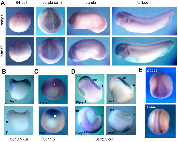
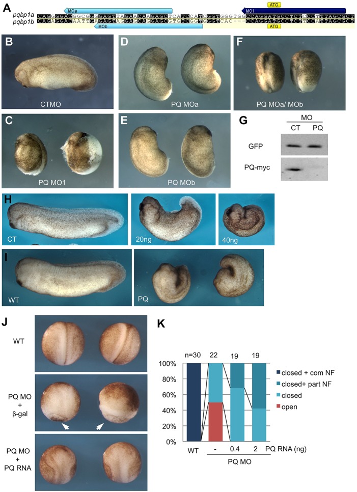
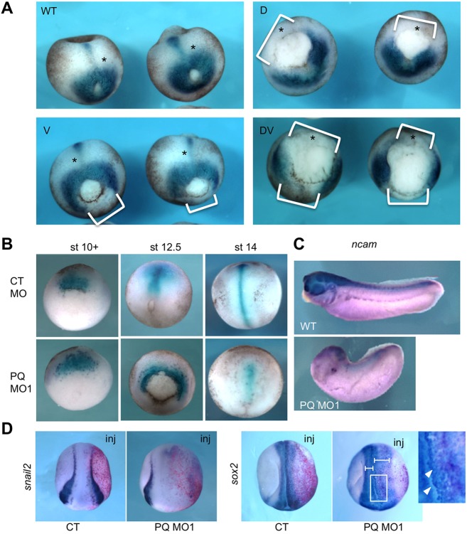
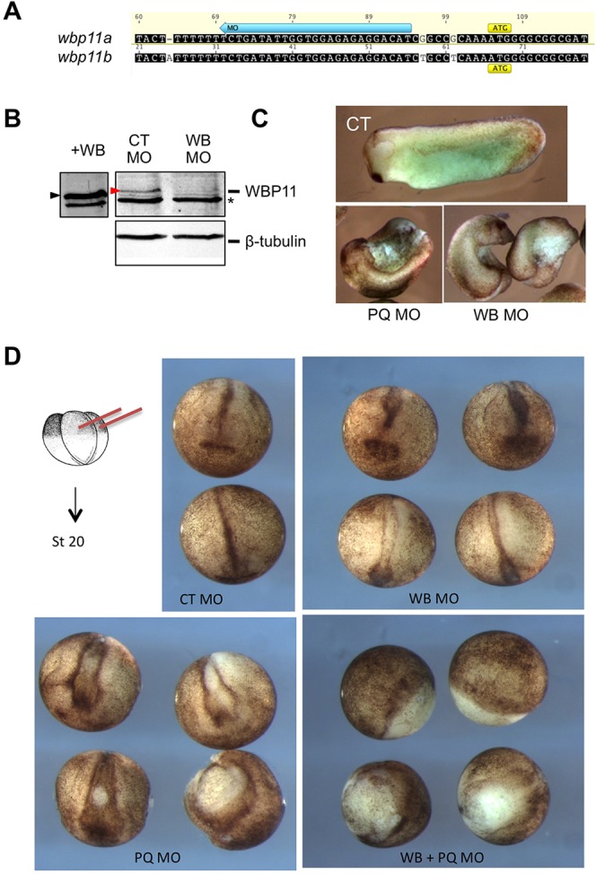
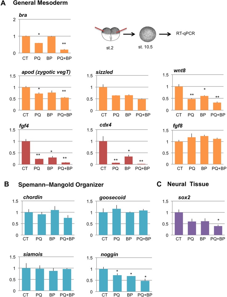
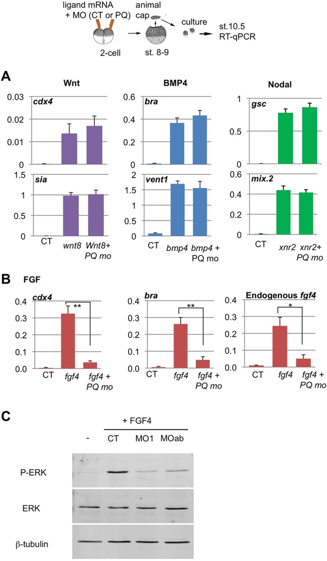
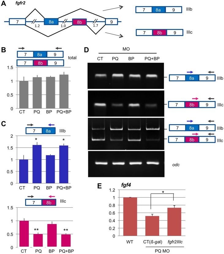
Alternative splicing of pre-mRNAs is an important means of regulating developmental processes, yet the molecular mechanisms governing alternative splicing in embryonic contexts are just beginning to emerge. Polyglutamine-binding protein 1 (PQBP1) is an RNA-splicing factor that, when mutated, in humans causes Renpenning syndrome, an X-linked intellectual disability disease characterized by severe cognitive impairment, but also by physical defects that suggest PQBP1 has broader functions in embryonic development. Here, we reveal essential roles for PQBP1 and a binding partner, WBP11, in early development of Xenopus embryos. Both genes are expressed in the nascent mesoderm and neurectoderm, and morpholino knockdown of either causes defects in differentiation and morphogenesis of the mesoderm and neural plate. At the molecular level, knockdown of PQBP1 in Xenopus animal cap explants inhibits target gene induction by FGF but not by BMP, Nodal or Wnt ligands, and knockdown of either PQBP1 or WBP11 in embryos inhibits expression of fgf4 and FGF4-responsive cdx4 genes. Furthermore, PQBP1 knockdown changes the alternative splicing of FGF receptor-2 (FGFR2) transcripts, altering the incorporation of cassette exons that generate receptor variants (FGFR2 IIIb or IIIc) with different ligand specificities. Our findings may inform studies into the mechanisms underlying Renpenning syndrome.

Keywords: Alternative splicing; FGF; FGF receptor; Mesoderm; Neural; PQBP1; Renpenning syndrome; WBP11; Xenopus.


Figures

Fig. 1.
Fig. 1.
pqbp1 and wbp11 genes are similarly expressed in mesoderm and neural tissues during Xenopus development. (A) WISH detection of pqbp1 and wbp11 transcripts in 64-cell blastula, neurula and tailbud tadpole; lateral views except anterior (ant) view of neurula. (B-E) WISH on intact (C,E) or longitudinally bisected (B,D) embryos with probes indicated. Asterisks mark the dorsal blastopore lip. (B) pqbp1 transcripts are present in the animal pole ectoderm and marginal zone in early gastrula (stage 10.5), upper panel; sense probe, lower panel. (C) Expression of pqbp1 and chordin in gastrulae, stage 11.5. (D) Expression of pqbp1, wbp11, chordin and sox2 in late gastrulae, dorsal-posterior views. Expression of pqbp1 overlaps with chordin in dorsal/axial mesoderm (arrows) and with sox2 in anterior neurectoderm. (E) Expression of pqbp1 and ncam mRNA in the neural plate of early neurula embryos. Note pqbp1 expression is in a broader region than ncam. an, anterior neurectoderm; chd, chordin; S, pqbp1 sense probe.
Fig. 2.
Fig. 2.
Knockdown of PQBP1 causes defective embryo morphogenesis. (A) Design of three pqbp1 antisense morpholino oligonucleotides (MOs). MO1 (purple bar) targets the ATG start codon of pqbp1a and pqbp1b (with three nucleotide mismatches), while MOa and MOb (blue bars) target the 5′ UTRs of pqbp1 a and b, respectively. Identical nucleotides between pqbp1 a and b are indicated by black background. (B-F) Tailbud tadpole stage embryos injected bilaterally at the two-cell stage with 50 ng control (CT) or pqbp1 (PQ) MOs as indicated (embryos in F received 100 ng MOs in total). (G) Translation of C-terminal myc-tagged Xenopus PQBP1 (PQ-myc) was blocked by co-injection of pqbp1 MO1 but not control MO (CT). GFP mRNA co-injected as a negative control for MO targeting and loading control. (H) Phenotypes of tailbud stage embryos dorsally targeted with control (CT) or the indicated amount of pqbp1 MO1. (I) Overexpression of PQBP1 via injected mRNA (PQ) perturbs normal development. Left panel, wild-type embryo (stage 26); right panel, embryos injected dorsally with pqbp1 mRNA (2 ng) at the four-cell stage. (J,K) Gastrulation and neurulation defects are partially rescued by MO-resistant pqbp1 mRNA. PQ MO (30 ng) co-injected dorsally with 2 ng lacZ mRNA encoding β-galactosidase (β-gal), displayed perturbed gastrulation (arrowheads point to open blastopores) substantially rescued by co-injection of morpholino-resistant pqbp1 (2 ng) (J). Embryos injected with PQ MO and either 0.4 or 2 ng of pqbp1 mRNA scored for the following phenotypes (K): closed blastopore with complete neural folds (closed+com NF), closed blastopore with partial neural folds (closed+part NF), closed blastopore without neural folds (closed) or open blastopore (open). WT, wild type.
Fig. 3.
Fig. 3.
The effects of PQBP1 knockdown on embryonic mesoderm and neurectoderm. (A) Expression of the pan-mesodermal marker brachyury (bra) in late gastrula (stage 12.5) embryos injected with 20 ng pqbp1 MO1 into dorsal (D), ventral (V) or both (DV) blastomeres at the four-cell stage. Note lack of bra expression wherever the pqbp1 MO was injected (white brackets). (B) Expression of the dorsal (axial) mesoderm marker chordin in embryos injected dorsally with 20 ng of either control MO (CT MO) or pqbp1 MO1 (PQ MO1) at the four-cell stage. (C) Expression of neural marker ncam in uninjected (WT) or pqbp1 MO1-injected embryos targeted dorsally as in B. Lateral views with anterior to left. (D) Expression of the neural crest marker snail2 (slug) and the neural marker sox2 in neurula embryos (stage 19) injected in a single two-cell stage blastomere with 20 ng pqbp1 MO1 or control MO (CT) along with β-galactosidase lineage tracer (red). Dorsal views with anterior down. Perturbed neural folding is shown by differences between width of left and right neural folds (brackets). Loosely adherent sox2-positive cells, marked by arrowheads in the magnified view of the boxed area. WT, wild-type control embryos.
Fig. 4.
Fig. 4.
WBP11 knockdown resembles and enhances PQBP1 knockdown phenotypes. (A) WBP11 MO (blue arrow) targets the 5′ UTR of both X. laevis wbp11 homeologs. (B) Expression of endogenous WBP11 was blocked by injection of 40 ng wbp11 MO (WB MO) but not control MO (CT MO). Endogenous (red arrowhead) and overexpressed (black arrowhead) WBP11 were detected by western blot with anti-WBP11 antibodies. An asterisk indicates a non-specific band that, along with β-tubulin staining, controls for sample loading. (C) Tailbud stage embryos injected into two dorsal cells at the four-cell stage with 100 ng control MO (CT), a mix of pqbp1 MOa and MOb (50 ng each; PQ MO) or 50 ng wbp11 MO (WB MO). (D) Neurula (stage 20, anterior view) embryos injected dorsally at the four-cell stage (schematic drawing) with 75 ng control MO (CT), 25 ng wbp11 MO (WB), a mixture of pqbp1 MOa plus MOb (25 ng each; 50 ng total; PQ) or a combination of wbp11 and pqbp1 MOa and MOb (25 ng each; 75 ng total; PQ+WB).
Fig. 5.
Fig. 5.
Effects of PQBP1 and WBP11 knockdown on embryonic gene expression. qPCR performed on early gastrula (stage 10.5) morphant embryos injected bilaterally at the two-cell stage with 150 ng control (CT), 100 ng pqbp1 (PQ), 50 ng wbp11 (BP) or a combination of pqbp1 and wbp11 (PQ+BP) MOs (150 ng total). Values were plotted relative to the control cap signal and shown as mean±s.e.m of n=3 with Student's t-test to control embryos (CT) (*P<0.05 or **P<0.01). (A) Expression of general mesodermal markers brachyury (bra), antipodean (apod), sizzled, wnt8, fgf4, cdx4 and fgf8. (B) Expression of Spemann–Mangold organizer-specific markers, chordin, goosecoid, siamois and noggin. (C) Expression of early neural marker sox2.
Fig. 6.
Fig. 6.
Effects of PQBP1 knockdown on animal cap response to growth factors. (A,B) Two-cell embryos were injected into the animal pole with growth factor mRNAs and MOs, animal caps were cut at mid-blastula and harvested at the equivalent of early gastrula stage, followed by qPCR, as depicted (top). Results were analyzed and plotted as per Fig. 5. (A) Marker gene induction by Wnt8 (50 pg), BMP4 (500 pg) and Nodal2 (Xnr2; 100 pg) was not affected by PQBP1 knockdown (50 ng MO). There was no statistically significant difference between CT and PQBP1 MO-injected caps treated with each ligand (Student's t-test, n=3). (B) Marker gene induction by FGF4 was significantly reduced by PQBP1 knockdown (*P<0.05 or **P<0.01; Student's t-test, triplicate biological replicates). Animal caps were injected with fgf4 mRNA (1 pg) and either control MO (50 ng), pqbp1 MO1 (50 ng) or MOa+MOb (25 ng each). (C) Phosphorylation of MAPK (Erk) was induced in fgf4-injected animal caps, but blocked by co-injection of pqbp1 MO, either MO1 or MOa+MOb. The levels of β-tubulin and total MAPK protein did not change among these samples.
Fig. 7.
Fig. 7.
Alternative splicing pattern of fgfr2 exon 8a/b is altered by PQBP1 knockdown. (A) Alternative splicing incorporates either exon 8a or 8b into fgfr2 transcripts, which generates two isoforms of FGFR2: IIIb and IIIc, respectively (see text). Intron sizes (kb) indicated. (B,C) Sum total of qPCR measurements of fgfr2IIIb/c transcripts (B), and the relative levels of each splicing variant (C), on total RNA extracted from early gastrula (stage 10.5) morphant embryos bilaterally injected at the two-cell stage with MOs: 150 ng control (CT), 100 ng PQBP1 (PQ), 50 ng WBP11 (BP), or combined PQBP1 and WBP11 (PQ+BP) (150 ng total). Samples were prepared and analyzed as per Fig. 5 (*P<0.05 or **P<0.01; Student's t-test, n=3). (D) Semi-quantitative RT-PCR amplification products of alternative spliced exon 8a or 8b from embryos injected with morpholinos (as above). (E) Partial rescue of fgf4 expression in pqbp1 morphants (100 ng pqbp1 MO) by co-injection of 1 ng Xenopus fgfr2IIIc (*P<0.05; Student's t-test, n=3).

Similar articles

Cited by

References

    1. Alexandrova, E. M. and Thomsen, G. H. (2006). Smurf1 regulates neural patterning and folding in Xenopus embryos by antagonizing the BMP/Smad1 pathway. Dev. Biol. 299, 398-410 10.1016/j.ydbio.2006.08.009 - DOI - PMC - PubMed
    1. Amaya, E., Stein, P. A., Musci, T. J. and Kirschner, M. W. (1993). FGF signalling in the early specification of mesoderm in Xenopus. Development 118, 477-487. - PubMed
    1. Blunt, A. G., Lawshé, A., Cunningham, M. L., Seto, M. L., Ornitz, D. M. and MacArthur, C. A. (1997). Overlapping expression and redundant activation of mesenchymal fibroblast growth factor (FGF) receptors by alternatively spliced FGF-8 ligands. J. Biol. Chem. 272, 3733-3738 10.1074/jbc.272.6.3733 - DOI - PubMed
    1. Busch, A., Engemann, S., Lurz, R., Okazawa, H., Lehrach, H. and Wanker, E. E. (2003). Mutant huntingtin promotes the fibrillogenesis of wild-type huntingtin: a potential mechanism for loss of huntingtin function in Huntington's disease. J. Biol. Chem. 278, 41452-41461 10.1074/jbc.M303354200 - DOI - PubMed
    1. Callery, E. M., Smith, J. C and Thomsen, G. H. (2005). The ARID domain protein dril1 is necessary for TGF(beta) signaling in Xenopus embryos. Dev. Biol. 278, 542-559 10.1016/j.ydbio.2004.11.017 - DOI - PubMed

Publication types

MeSH terms