. 2014 Sep 11:2:132.
doi: 10.1186/s40478-014-0132-8.

New α- and γ-synuclein immunopathological lesions in human brain

New α- and γ-synuclein immunopathological lesions in human brain

Irina Surgucheva et al. Acta Neuropathol Commun. .

Abstract

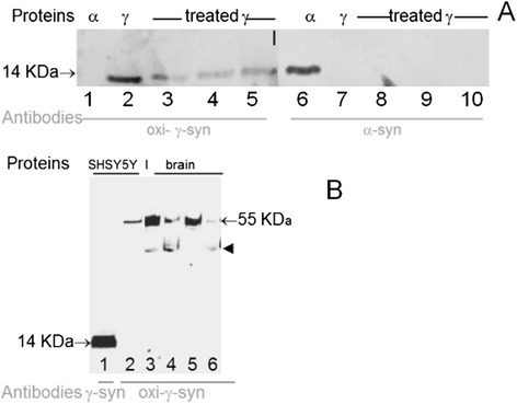
Introduction: Several neurodegenerative diseases are classified as proteopathies as they are associated with the aggregation of misfolded proteins. Synucleinopathies are a group of neurodegenerative disorders associated with abnormal deposition of synucleins. α-Synucleinopathies include Parkinson's disease, dementia with Lewy bodies, and multiple system atrophy. Recently accumulation of another member of the synuclein family- γ-synuclein in neurodegenerative diseases compelled the introduction of the term γ-synucleinopathy. The formation of aggregates and deposits of γ-synuclein is facilitated after its oxidation at methionine 38 (Met38).

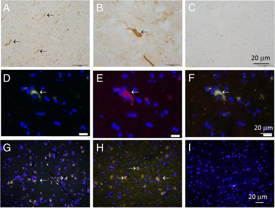
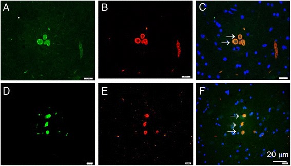
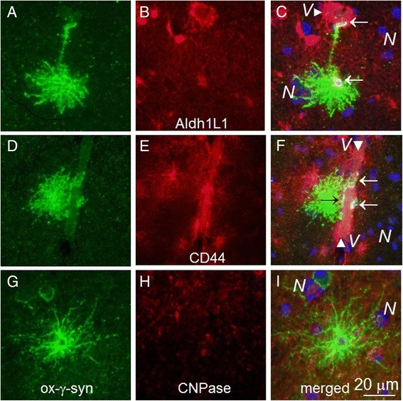
Results: Several types of intracytoplasmic inclusions containing post-translationally modified α- and γ-synucleins are detected. Oxidized Met38-γ-synuclein forms aberrant inclusions in amygdala and substantia nigra. Double staining revealed colocalization of oxidized-γ-synuclein with α-synuclein in the cytoplasm of neurons. Another type of synuclein positive inclusions in the amygdala of dementia with Lewy bodies patients has the appearance of Lewy bodies. These inclusions are immunoreactive when analyzed with antibodies to α-synuclein phosphorylated on serine 129, as well as with antibodies to oxidized-γ-synuclein. Some of these Lewy bodies have doughnut-like shape with round or elongated shape. The separate immunofluorescent images obtained with individual antibodies specific to oxidized-γ-synuclein and phospho-α-synuclein clearly shows the colocalization of these synuclein isoforms in substantia nigra inclusions. Phospho-α-synuclein is present almost exclusively at the periphery of these structures, whereas oxidized-γ-syn immunoreactivity is also located in the internal parts forming dot-like pattern of staining.

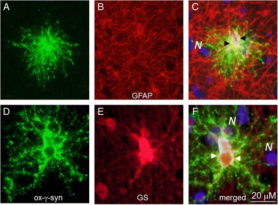
Conclusions: These results reveal new γ-synuclein positive lesions in human brain. Oxidized-γ-synuclein is colocalized with phospho-α-synuclein in doughnut-like inclusions. Several types of astrocytes with different morphology are immunopositive for oxidized-γ-synuclein.


Figures

Figure 1
Figure 1
Specificity of synuclein antibody toward recombinant proteins (A) and extracts from cell culture and brain (B). A: Recombinant α-syn (lanes 1 and 6), γ-syn (lanes 2 and 7) and γ-syn treated with DA (oxi-γ-syn, lanes 3–5 and 8–10) were separated in 12% polyacrylamide gel with SDSNa and tested with antibodies specific to different synucleins isoforms. The following amounts of protein were subjected to electrophoresis: lanes 2 and 7 – untreated -γ-syn - 62.5 ng. Since in the course of the treatment by 250 μM DA the amount of immunoreactive monomeric γ-syn was reduced, we applied increasing amounts of proteins: 100 ng on lanes 3 and 8; 187.5 ng on lanes 4 and 9; and 225 ng on lanes 5 and 10. WB was probed with antibodies against oxi-γ- syn (lanes 1–5) and antisheep-α-synuclein (lanes 6–10). B: Antibody to unmodified γ-syn (ABcam, 55424) recognizes monomeric form of γ-syn (lane 1), while antibody to oxi-γ-syn recognizes tetrameric γ-syn in cell extracts of SH-SY5Y cells overexpressing γ-syn (lane 2) and in extracts of human brain (3–6). Lanes 3 and 5 extracts from amygdala were analyzed; lanes 4 and 6 – extracts from substantia nigra from control individuals.
Figure 2
Figure 2
Colocalization of α-syn with oxi-γ-syn in amygdala and substantia nigra in AD with LB patients. Frozen (two top rows) and paraffin-embedded sections (bottom row) of substantia nigra and amygdala of healthy (A, G) and AD patients with DLB (H, I). The top row: frozen sections (A and B) were stained using H&R- DAB –procedure and oxi-γ-syn antibody. A- substantia nigra from K46 healthy individual, B - substantia nigra from S3 patient (AD with LB), C- the same as in A, but without primary antibody. In the middle row the sections of substantia nigra from S3 patient were stained by oxi-γ-syn antibody; D - α-syn antibody (Abcam ab6162, antisheep); E - oxi-γ-syn antibody; F - colocalization of both antibodies. The bottom row. The sections were double stained with antibody against α-syn (red, Abcam ab 6162, antisheep) and oxi-γ-syn (green, antirabbit, homemade antibody). Yellow cytoplasmic staining is shown by an arrow in a sample from amygdala of K21-healthy donor (G) and in a sample from amygdala taken from a patient S4 with AD with LB (H) substantia nigra from patient S3 suffering from mild AD with LB (I). Antibody to unmodified γ-syn (clone 1H10D2, Antagene) (red) reveals a weak staining of unmodified γ-syn in the cytoplasm of neurons.
Figure 3
Figure 3
The presence of post-translationally modified α- and γ-syn in LB. The immunofluorescence staining of sections by individual antibody. A-C - substantia nigra of S3 patient (AD with LB). A - oxi-γ-syn immunoreactivity, B- phospho-α-syn immunoreactivity, C- merged. D-F -LB from amygdala of a patient K33 with DLB. D - oxi-γ-syn immunoreactivity, E- phospho-α-syn immunoreactivity, F – merged. Blue - DAPI staining.
Figure 4
Figure 4
Oxi-γ-syn positive cells with long processes are identified as astrocytes with specific markers, GFAP (A-C) and glutamine synthetase (D- F). GS-positive astrocytes have relatively long branched processes (D) compared to more compact bushy-like spherical GFAP-positive astrocytes (A).
Figure 5
Figure 5
Oxi-γ-syn positive astrocytes have a partial colocalization with astrocyte markers Aldh1L1 (C) and CD44 (F) (arrows) and are not colocalized with an oligodendrocyte marker CNPase (I). A, D, G – oxi-γ-syn staining. B – Aldh1L1, E- CD44, H –CNPase staining. Some of oxi-γ-syn-positive astrocytes reside near large blood vessels ( V, C and F) with processes extending toward the vessel surface (C).

References

    1. Edison P, Rowe CC, Rinne JO, Ng S, Ahmed I, Kemppainen N, Villemagne VL, O’Keefe G, Någren K, Chaudhury KR, Masters CL, Brooks DJ. Amyloid load in Parkinson’s disease dementia and Lewy body dementia measured with [11C]PIB positron emission tomography. J Neurol Neurosurg Psychiatry. 2008;79(12):1331–1338. doi: 10.1136/jnnp.2007.127878. - DOI - PubMed
    1. Resende R, Marques SC, Ferreiro E, Simões I, Oliveira CR, Pereira CM. Effect of α-synuclein on amyloid β-induced toxicity: relevance to Lewy body variant of Alzheimer disease. Neurochem Res. 2013;38(4):797–806. doi: 10.1007/s11064-013-0982-7. - DOI - PubMed
    1. Hamilton RL. Lewy bodies in Alzheimer’s disease: a neuropathological review of 145 cases using alpha-synuclein immunohistochemistry. Brain Pathol. 2000;10:378–384. doi: 10.1111/j.1750-3639.2000.tb00269.x. - DOI - PMC - PubMed
    1. Pronin AN, Morris AJ, Surguchov A, Benovic JL. Synucleins are a novel class of substrates for G protein-coupled receptor kinases. J Biol Chem. 2000;275(34):26515–26522. doi: 10.1074/jbc.M003542200. - DOI - PubMed
    1. Foulds PG, Mitchell JD, Parker A, Turner R, Green G, Diggle P, Hasegawa M, Taylor M, Mann D, Allsop D. Phosphorylated α-synuclein can be detected in blood plasma and is potentially a useful biomarker for Parkinson’s disease. FASEB J. 2011;25(12):4127–4137. doi: 10.1096/fj.10-179192. - DOI - PubMed

Publication types