

Cocaine potentiates cathepsin B secretion and neuronal apoptosis from HIV-infected macrophages
- PMID: 25209871
- PMCID: PMC4209444
- DOI: 10.1007/s11481-014-9563-z
Cocaine potentiates cathepsin B secretion and neuronal apoptosis from HIV-infected macrophages
Abstract
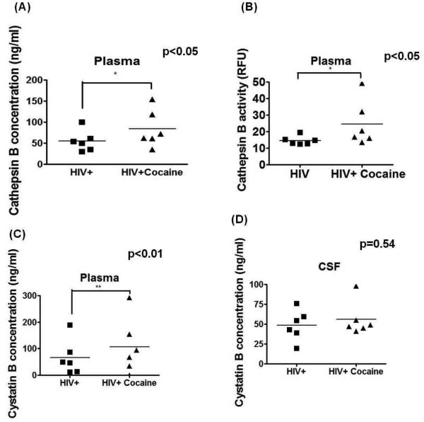
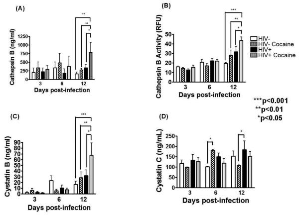
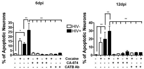
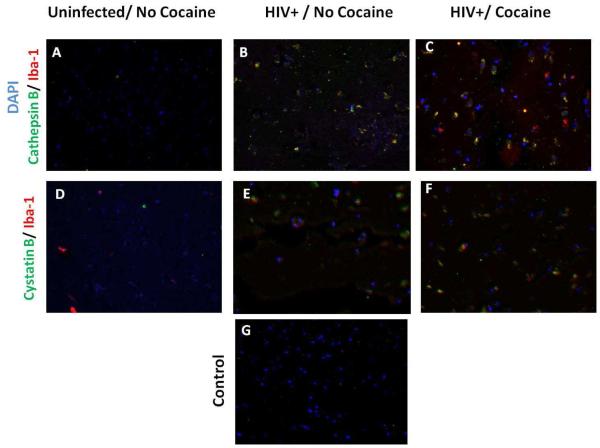
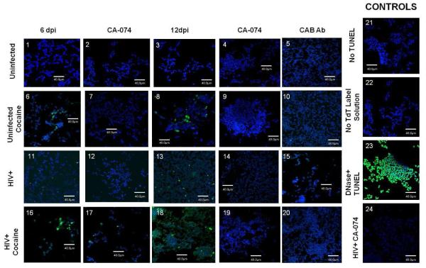
Substance abuse is a risk factor for HIV infection and progression to AIDS. Recent evidence establishes that cocaine use promotes brain perivascular macrophage infiltration and microglia activation. The lysosomal protease cathepsin B is increased in monocytes from patients with HIV dementia and its secretion induces 10-15% of neurotoxicity. Here we asked if cocaine potentiates cathepsin B secretion from HIV-infected monocyte-derived macrophages (MDM) and its effect in neuronal apoptosis. Samples of plasma, CSF, and post-mortem brain tissue from HIV positive patients that used cocaine were tested for cathepsin B and its inhibitors to determine the in vivo relevance of these findings. MDM were inoculated with HIV-1ADA, exposed to cocaine, and the levels of secreted and bioactive cathepsin B and its inhibitors were measured at different time-points. Cathepsin B expression (p < 0.001) and activity (p < 0.05) increased in supernatants from HIV-infected cocaine treated MDM compared with HIV-infected cocaine negative controls. Increased levels of cystatin B expression was also found in supernatants from HIV-cocaine treated MDM (p < 0.05). A significant increase in 30% of apoptotic neurons was obtained that decreased to 5% with the specific cathepsin B inhibitor (CA-074) or with cathepsin B antibody. Cathepsin B was significantly increased in the plasma and post-mortem brain tissue of HIV/cocaine users over non-drug users. Our results demonstrated that cocaine potentiates cathepsin B secretion in HIV-infected MDM and increase neuronal apoptosis. These findings provide new evidence that cocaine synergize with HIV-1 infection in increasing cathepsin B secretion and neurotoxicity.
Figures
References
-
- Bansal AK, Mactutus CF, Nath A, et al. Neurotoxicity of HIV-1 proteins gp120 and Tat in the rat striatum. Brain research. 2000;879:42–9. - PubMed
-
- Brix K, Dunkhorst A, Mayer K, Jordans S. Cysteine cathepsins: cellular roadmap to different functions. Biochimie. 2008;90:194–207. doi: 10.1016/j.biochi.2007.07.024. - PubMed
-
- Cho K, Yoon SY, Choi JE, et al. CA-074Me, a cathepsin B inhibitor, decreases APP accumulation and protects primary rat cortical neurons treated with okadaic acid. Neuroscience letters. 2013;548:222–7. doi: 10.1016/j.neulet.2013.05.056. - PubMed
Publication types
MeSH terms
Substances
Grants and funding
- R24 MH059724/MH/NIMH NIH HHS/United States
- U01 MH083500/MH/NIMH NIH HHS/United States
- R24 NS045491/NS/NINDS NIH HHS/United States
- 1-U54NS431/NS/NINDS NIH HHS/United States
- P20 RR016470/RR/NCRR NIH HHS/United States
- U01MH083501/MH/NIMH NIH HHS/United States
- U01 MH083501/MH/NIMH NIH HHS/United States
- U01MH083545/MH/NIMH NIH HHS/United States
- 8G12-MD007600/MD/NIMHD NIH HHS/United States
- U24 MH100929/MH/NIMH NIH HHS/United States
- U01 MH083507/MH/NIMH NIH HHS/United States
- U24 MH100931/MH/NIMH NIH HHS/United States
- R01MH083516/MH/NIMH NIH HHS/United States
- R25 GM061838/GM/NIGMS NIH HHS/United States
- U24 MH100928/MH/NIMH NIH HHS/United States
- 5U01MH083500/MH/NIMH NIH HHS/United States
- R24MH59724/MH/NIMH NIH HHS/United States
- R24 MH059745/MH/NIMH NIH HHS/United States
- R24MH59745/MH/NIMH NIH HHS/United States
- U54 NS043011/NS/NINDS NIH HHS/United States
- U54NS043011/NS/NINDS NIH HHS/United States
- P20RR016470-12/RR/NCRR NIH HHS/United States
- R24 NS038841/NS/NINDS NIH HHS/United States
- R24 NS45491/NS/NINDS NIH HHS/United States
- NS 38841/NS/NINDS NIH HHS/United States
- R25GM061838/GM/NIGMS NIH HHS/United States
- G12 RR003051/RR/NCRR NIH HHS/United States
- N01 MH032002/MH/NIMH NIH HHS/United States
- U01MH083506/MH/NIMH NIH HHS/United States
- U01 MH083545/MH/NIMH NIH HHS/United States
- U01 MH083506/MH/NIMH NIH HHS/United States
- R01 MH083516/MH/NIMH NIH HHS/United States
- U01MH083507/MH/NIMH NIH HHS/United States
- G12 MD007600/MD/NIMHD NIH HHS/United States
LinkOut - more resources
Full Text Sources
Other Literature Sources
Medical
Research Materials
Miscellaneous
