Review
. 2014 Oct 31;289(44):30205-30212.
doi: 10.1074/jbc.R114.565515. Epub 2014 Sep 10.

Evolutionary aspects of enzyme dynamics

Affiliations
Review

Evolutionary aspects of enzyme dynamics

Judith P Klinman et al. J Biol Chem. .

Abstract

The role of evolutionary pressure on the chemical step catalyzed by enzymes is somewhat enigmatic, in part because chemistry is not rate-limiting for many optimized systems. Herein, we present studies that examine various aspects of the evolutionary relationship between protein dynamics and the chemical step in two paradigmatic enzyme families, dihydrofolate reductases and alcohol dehydrogenases. Molecular details of both convergent and divergent evolution are beginning to emerge. The findings suggest that protein dynamics across an entire enzyme can play a role in adaptation to differing physiological conditions. The growing tool kit of kinetics, kinetic isotope effects, molecular biology, biophysics, and bioinformatics provides means to link evolutionary changes in structure-dynamics function to the vibrational and conformational states of each protein.

Keywords: Alcohol Dehydrogenase (ADH); Enzyme; Enzyme Catalysis; Enzyme Kinetics; Enzyme Mechanism; Enzyme Mutation; Evolution; Folate Metabolism; Molecular Evolution; Protein Evolution.


Figures

FIGURE 1.
FIGURE 1.
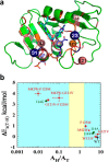
Structural, genetic, and functional features of DHFR. a, DHFR structure (Protein Data Bank (PDB) ID 1rx2) colored based on the genetic coupling analysis as Conserved (red), Strongly Coupled, (pink), and Weakly Coupled, (orange). The nicotinamide cofactor and folate are highlighted as blue and light blue sticks, respectively, and a black arrow is drawn at the location of the C–H→C transfer (between C4 of the nicotinamide to C6 of the pterin). Highlighted as spheres with the same color code are the α-carbons of the four coevolving residues that are discussed in the text above. Highlighted as dark blue spheres are Asn-23 and Gly-51, which are the sites of the evolution-induced insertions discussed below. b, correlation of temperature dependence parameters of T-KIEint for WT (black), distal (red), and active site Ile-14 (green) mutants of ecDHFR, where error bars represent S.D. The yellow block represents semi-classical range of Arrhenius pre-exponential factor (0.3–1.7) (46). Reproduced from Ref. . with permission from the American Chemical Society.
FIGURE 2.
FIGURE 2.
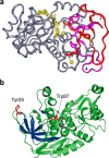
Structural features of ht-ADH. a, the five peptides (1–4 and 7) that increase their flexibility above 30 °C in ht-ADH reside within the substrate-binding site and are colored orange and fuchsia. The cofactor NAD+ has been modeled into the active site and is colored yellow, as are the catalytic zinc ion (near the nicotinamide ring of cofactor) and the structural zinc ion. Reproduced with permission from (37), copyright (2004) National Academy of Sciences, U.S.A. b, the relationship of the surface Tyr-25 (red) to the active site Trp-87 (red). Bound substrate, adjacent to Trp-87, is black. The series of β-sheets that increase their flexibility above 30 °C (see a above) are colored dark blue.
FIGURE 3.
FIGURE 3.
A diagram illustrating the distribution of conformational sub-states as a function of temperature in ht-ADH. Reproduced with permission from Ref. , copyright (2011) National Academy of Sciences, U.S.A. a–c indicate the redistribution of enzyme from an inactive region of the conformational landscape (state 4) into the regions that can support hydride transfer (states 1-3) as the temperature is raised above 30 °C. The mathematical model treats the rate as a function of a temperature-dependent equilibration, Keq, and the rate constant, kintrinsic. d shows the ability to achieve Aobserved = 1025 s−1 via a compensatory increase in ΔH and TΔS. The extrapolation to Aintrinsic is shown by the dashed line, and the extrapolation to Aobserved is shown by the solid line.

Similar articles

Cited by

References

    1. Süel G. M., Lockless S. W., Wall M. A., Ranganathan R. (2003) Evolutionarily conserved networks of residues mediate allosteric communication in proteins. Nat. Struct. Biol. 10, 59–69 - PubMed
    1. Hammes-Schiffer S., Benkovic S. J. (2006) Relating protein motion to catalysis. Annu. Rev. Biochem. 75, 519–541 - PubMed
    1. Radkiewicz J. L., Brooks C. L. (2000) Protein dynamics in enzymatic catalysis: exploration of dihydrofolate reductase. J. Am. Chem. Soc. 122, 225–231
    1. Agarwal P. K., Billeter S. R., Rajagopalan P. T. R., Benkovic S. J., Hammes-Schiffer S. (2002) Network of coupled promoting motions in enzyme catalysis. Proc. Natl. Acad. Sci. U.S.A. 99, 2794–2799 - PMC - PubMed
    1. Rod T. H., Radkiewicz J. L., Brooks C. L. (2003) Correlated motion and the effect of distal mutations in dihydrofolate reductase. Proc. Natl. Acad. Sci. U.S.A. 100, 6980–6985 - PMC - PubMed

Publication types

Substances

LinkOut - more resources