. 2014 Oct 24;289(43):29859-73.
doi: 10.1074/jbc.M114.591073. Epub 2014 Sep 10.

Human 2-oxoglutarate dehydrogenase complex E1 component forms a thiamin-derived radical by aerobic oxidation of the enamine intermediate

Affiliations

Human 2-oxoglutarate dehydrogenase complex E1 component forms a thiamin-derived radical by aerobic oxidation of the enamine intermediate

Natalia S Nemeria et al. J Biol Chem. .

Abstract

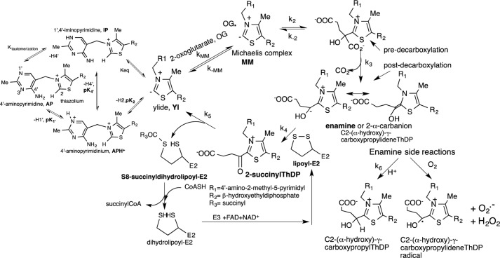
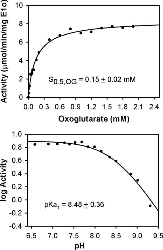
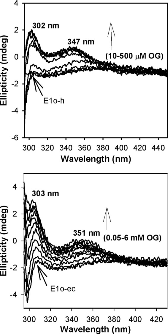
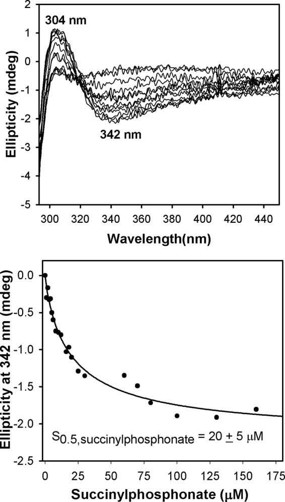
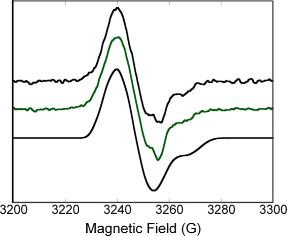
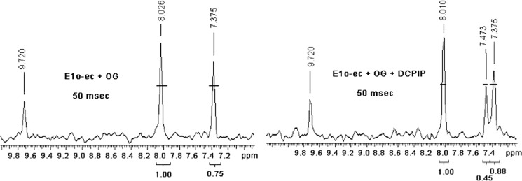
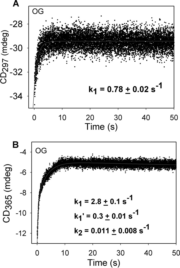
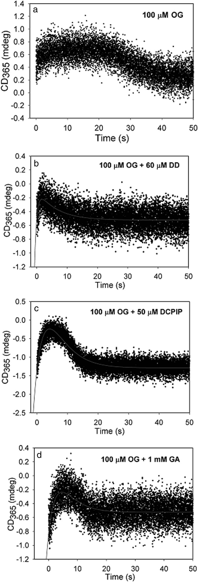
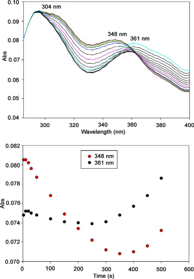
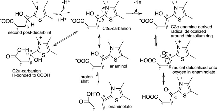
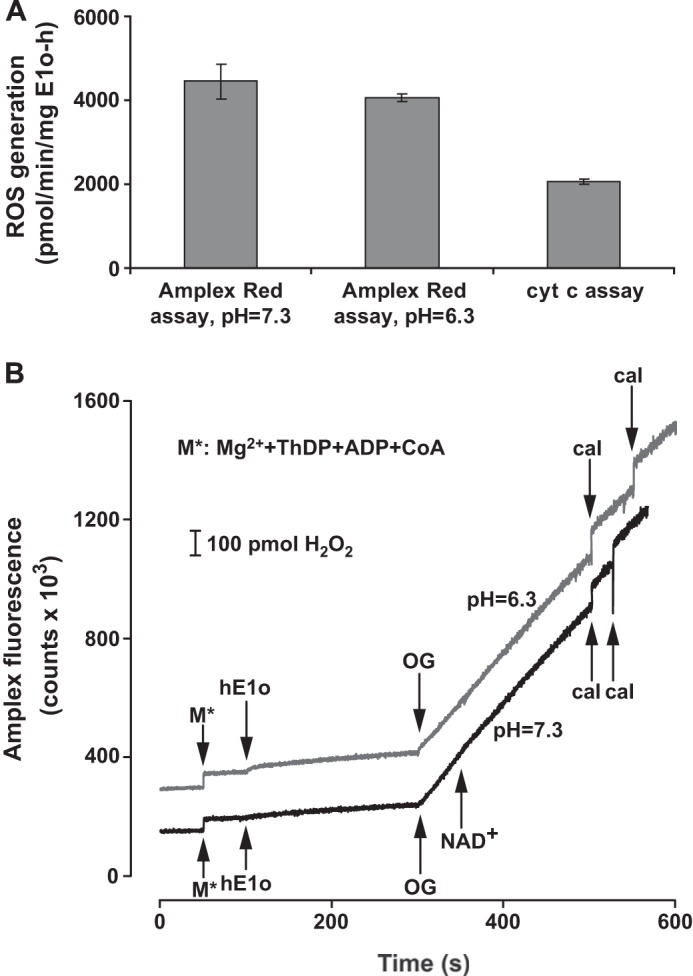
Herein are reported unique properties of the human 2-oxoglutarate dehydrogenase multienzyme complex (OGDHc), a rate-limiting enzyme in the Krebs (citric acid) cycle. (a) Functionally competent 2-oxoglutarate dehydrogenase (E1o-h) and dihydrolipoyl succinyltransferase components have been expressed according to kinetic and spectroscopic evidence. (b) A stable free radical, consistent with the C2-(C2α-hydroxy)-γ-carboxypropylidene thiamin diphosphate (ThDP) cation radical was detected by electron spin resonance upon reaction of the E1o-h with 2-oxoglutarate (OG) by itself or when assembled from individual components into OGDHc. (c) An unusual stability of the E1o-h-bound C2-(2α-hydroxy)-γ-carboxypropylidene thiamin diphosphate (the "ThDP-enamine"/C2α-carbanion, the first postdecarboxylation intermediate) was observed, probably stabilized by the 5-carboxyl group of OG, not reported before. (d) The reaction of OG with the E1o-h gave rise to superoxide anion and hydrogen peroxide (reactive oxygen species (ROS)). (e) The relatively stable enzyme-bound enamine is the likely substrate for oxidation by O2, leading to the superoxide anion radical (in d) and the radical (in b). (f) The specific activity assessed for ROS formation compared with the NADH (overall complex) activity, as well as the fraction of radical intermediate occupying active centers of E1o-h are consistent with each other and indicate that radical/ROS formation is an "off-pathway" side reaction comprising less than 1% of the "on-pathway" reactivity. However, the nearly ubiquitous presence of OGDHc in human tissues, including the brain, makes these findings of considerable importance in human metabolism and perhaps disease.

Keywords: Dehydrogenase; Electron Paramagnetic Resonance (EPR); Mass Spectrometry (MS); Reactive Oxygen Species (ROS); Thiamin.


Figures

SCHEME 1.
SCHEME 1.
Mechanism of OGDHc with alternative fates of the enamine.
REACTION 1
REACTION 1
FIGURE 1.
FIGURE 1.
Kinetic parameters for human OGDHc. Top, dependence of the OGDHc activity on concentration of OG. The OGDHc was assembled from E1o-h, E2o-h, and E3 components at a mass ratio (μg/μg/μg) of 1:5:5, corresponding to the following concentrations of subunits: E1o-h (0.046 μm), E2o-h (0.60 μm), and E3 (0.52 μm). For experimental details, see “Experimental Procedures.” Data were fitted to a Hill equation (vo = vomax[OG]n/(S0.5n + [OG]n), and the trace is the regression fit line. nH = 0.92 ± 0.08. Bottom, pH dependence of OGDHc activity. The reaction was carried out in 0.1 m Tris·HCl in the pH range of 6.5–9.3. For details on OGDHc assembly and activity measurement, see “Experimental Procedures.” The values of activity were plotted to a curve defined by one ionizing group according to log(activity) = log(activitymax) − log(1 + 10(x − pKa1)), where x is the value of pH.
FIGURE 2.
FIGURE 2.
Circular dichroism titration of E1o-h and E1o-ec by OG. Top, E1o-h (18 μm active center concentration) in 50 mm KH2PO4 (pH 7.5) containing 0.20 mm ThDP and 1.0 mm MgCl2 was titrated by OG (10–500 μm) at 4 °C. Bottom, E1o-ec (20 μm active center concentration) was titrated by OG (0.05–6 mm) in 20 mm KH2PO4 (pH 7.0) containing 150 mm NaCl, 0.2 mm ThDP, and 1 mm MgCl2 at 20 °C.
FIGURE 3.
FIGURE 3.
Circular dichroism titration of E1o-h by succinylphosphonate. Top, the E1o-h (1.68 mg/ml; concentration of active centers, 14.9 μm) in 20 mm KH2PO4 (pH 7.5) containing 0.2 mm ThDP and 2.0 mm MgCl2 was titrated by succinylphosphonate (1–130 μm). Two CD bands observed could be assigned to 1′,4′-iminotautomer of the first predecarboxylation intermediate (positive CD band at 304 nm) and to the Michaelis complex (negative CD band at 342 nm). Bottom, the dependence of the CD at 342 nm on concentration of succinylphosphonate, leading to S0.5,succinylphosphonate of 20 ± 5 μm. Data were fitted to a Hill equation (CDo = CDomax[SP]n/(S0.5n + [SP]n), and the trace is the regression fit line. nH = 0.86 ± 0.11.
FIGURE 4.
FIGURE 4.
X-band (9 GHz) EPR spectra of the radical species generated in the E1o-h (top) and in the human oxoglutarate dehydrogenase complex (middle) along with a spectral simulation (bottom). Top, E1o-h (concentration of active centers = 0.449 mm) in 0.4 ml of 50 mm HEPES (pH 7.5) containing 0.15 m NaCl, 0.50 mm ThDP, and 1.0 mm MgCl2 was mixed with 10 mm OG at room temperature. The mixture was immediately transferred into an EPR tube and was flash-frozen in liquid nitrogen within 40 s of mixing all components. Middle, E1o-h, E2o-h, and E3 components were mixed at a concentration of subunits of 0.222 mm for each of the three components in 50 mm HEPES (pH 7.5) containing 0.50 mm ThDP, 2.0 mm MgCl2, and 0.15 m NaCl. The reaction was started by the addition of 10 mm OG, and sample was treated the same way as E1o-h above. Parameters used for the acquisition of the experimental spectra are presented under “Experimental Procedures.” Bottom, the parameters used for the simulation are consistent with the ThDP-enamine radical species (for details, see “Experimental Procedures”).
FIGURE 5.
FIGURE 5.
Distribution of ThDP-bound covalent intermediates in the reaction of E1o-ec. gCHSQC NMR spectra of the supernatant after acid quench of the E1o-ec reaction and removal of protein from the reaction. The [C2,C6′-13C2]ThDP-labeled E1o-ec was incubated with OG for 50 ms in the absence (left) and in the presence of DCPIP (right). The C6′H chemical shift of 8.01, 7.37, and 7.47 ppm corresponds to ThDP, C2-(α-hydroxy)-γ-carboxypropyl-ThDP, and succinyl-ThDP, respectively. The 7.36–7.37 ppm resonance has contributions from both species.
FIGURE 6.
FIGURE 6.
Pre-steady-state kinetics of E1o-ec with OG. A, time dependence of build-up of the C2-(α-hydroxy)-γ-carboxypropyl-ThDP intermediate (second postdecarboxylation intermediate in Scheme 1). The data points were fit using a single exponential equation (CD297(t) = CD1·ek1t + c), and the line is a regression fit trace. B, time dependence of enamine formation on E1o-ec. The data were fitted using the triple exponential equation (CDλ(t) = CD1·ek1t + CD2·ek1′t − CD2·ek2t + c), and the line is the regression fit trace. k1, k−1, and k2 are the apparent rate constants; c and CDmaxλ in the exponential rise to maximum model. All stopped-flow CD experiments were performed in 20 mm KH2PO4 (pH 7.0) with 0.5 mm ThDP and 2 mm MgCl2 at 20 °C.
FIGURE 7.
FIGURE 7.
Effect of different enamine acceptors on the CD at 365 nm. a, stopped-flow CD behavior at 365 nm, where E1o-ec from one syringe was mixed with 100 μm OG from the second syringe at 20 °C. In the other traces, E1o (38 μm active site concentration) from one syringe was mixed with the enamine acceptor (concentration indicated in the figure) premixed with OG in the second syringe. b, E2o-ec(1–176) didomain (DD). c, DCPIP. d, glyoxylate.
FIGURE 8.
FIGURE 8.
Reaction of E1o-ec with OG monitored by stopped-flow PDA. Top, E1o-ec spectra with 10 mm OG showing formation of ThDP-enamines, perhaps in different conformations/configurations. E1o-ec (40 μm concentration of active centers) was mixed with an equal volume of 10 mm OG in the same buffer at 25 °C. The reaction was monitored in the 280–400-nm wavelength range for 500 s. Bottom, time course of the formation of ThDP-enamines at the indicated wavelength.
SCHEME 2.
SCHEME 2.
Possible role of side chain carboxylate in stabilizing ThDP-bound enamine and radical.
FIGURE 9.
FIGURE 9.
Superoxide/H2O2 production by E1o-h. A, superoxide/H2O2 production by E1o-h as measured by the cyt c or the Amplex Red assay and expressed as the amount of superoxide or H2O2 produced (pmol)·min−1·mg E1o-h−1. Linear fitting to initial velocity data points generated always an R2 > 0.982 in the cyt c assay or greater in the Amplex Red assay. The primary data were corrected with the average slopes of the blanks (for both assays). Error bars, S.E. values for five (cyt c assay) or three (Amplex Red assay) parallel measurements. No statistically significant difference was found between the rates of H2O2 production measured at two different pH values by the Amplex Red assay. For experimental conditions, see “Experimental Procedures.” B, representative Amplex Red fluorescence traces for H2O2 generation by E1o-h at different pH values with additions indicated. For experimental conditions, see “Experimental Procedures.” Curves have been offset for clarity. For calibration of all experiments (designated as cal at the bottom), 100 pmol of H2O2 was applied in duplicates. 1 mm NAD+ was added to the reaction assay at pH 7.3.

References

    1. Perham R. N. (2000) Swinging arms and swinging domains in multifunctional enzymes: catalytic machines for multistep reactions. Annu. Rev. Biochem. 69, 961–1004 - PubMed
    1. Reed L. J. (2001) A trial of research from lipoic acid to α-keto acid dehydrogenase complexes. J. Biol. Chem. 276, 38329–38336 - PubMed
    1. Perham R. N. (1991) Domains, motifs, and linkers in 2-oxo acid dehydrogenase multienzyme complexes: a paradigm in the design of a multifunctional protein. Biochemistry 30, 8501–8512 - PubMed
    1. Gibson G. E., Park L. C. H., Sheu K. F. R., Blass J. P., Calingasan N. Y. (2000) The α-ketoglutarate dehydrogenase complex in neurodegeneration. Neurochem. Int. 36, 97–112 - PubMed
    1. Starkov A. A., Fiskum G., Chinopoulos C., Lorenzo B. J., Browne S. E., Patel M. S., Beal M. F. (2004) Mitochondrial α-ketoglutarate dehydrogenase complex generates reactive oxygen species. J. Neurosci. 24, 7779–7788 - PMC - PubMed

Publication types

LinkOut - more resources