

PCR-based detection of Toxoplasma gondii DNA in blood and ocular samples for diagnosis of ocular toxoplasmosis
- PMID: 25210066
- PMCID: PMC4313235
- DOI: 10.1128/JCM.01793-14
PCR-based detection of Toxoplasma gondii DNA in blood and ocular samples for diagnosis of ocular toxoplasmosis
Abstract
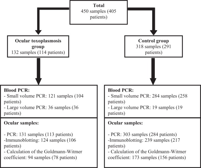
PCR detection of Toxoplasma gondii in blood has been suggested as a possibly efficient method for the diagnosis of ocular toxoplasmosis (OT) and furthermore for genotyping the strain involved in the disease. To assess this hypothesis, we performed PCR with 121 peripheral blood samples from 104 patients showing clinical and/or biological evidence of ocular toxoplasmosis and from 284 (258 patients) controls. We tested 2 different extraction protocols, using either 200 μl (small volume) or 2 ml (large volume) of whole blood. Sensitivity was poor, i.e., 4.1% and 25% for the small- and large-volume extractions, respectively. In comparison, PCR with ocular samples yielded 35.9% sensitivity, while immunoblotting and calculation of the Goldmann-Witmer coefficient yielded 47.6% and 72.3% sensitivities, respectively. Performing these three methods together provided 89.4% sensitivity. Whatever the origin of the sample (ocular or blood), PCR provided higher sensitivity for immunocompromised patients than for their immunocompetent counterparts. Consequently, PCR detection of Toxoplasma gondii in blood samples cannot currently be considered a sufficient tool for the diagnosis of OT, and ocular sampling remains necessary for the biological diagnosis of OT.
Copyright © 2014, American Society for Microbiology. All Rights Reserved.
Figures
Similar articles
-
Value of PCR for detection of Toxoplasma gondii in aqueous humor and blood samples from immunocompetent patients with ocular toxoplasmosis.J Clin Microbiol. 1999 Nov;37(11):3465-8. doi: 10.1128/JCM.37.11.3465-3468.1999. J Clin Microbiol. 1999. PMID: 10523535 Free PMC article.
-
Comparison of immunoblotting, calculation of the Goldmann-Witmer coefficient, and real-time PCR using aqueous humor samples for diagnosis of ocular toxoplasmosis.J Clin Microbiol. 2008 Jun;46(6):1965-7. doi: 10.1128/JCM.01900-07. Epub 2008 Apr 9. J Clin Microbiol. 2008. PMID: 18400917 Free PMC article.
-
Contributions of immunoblotting, real-time PCR, and the Goldmann-Witmer coefficient to diagnosis of atypical toxoplasmic retinochoroiditis.J Clin Microbiol. 2009 Jul;47(7):2131-5. doi: 10.1128/JCM.00128-09. Epub 2009 May 13. J Clin Microbiol. 2009. PMID: 19439541 Free PMC article.
-
Acquired Ocular Toxoplasmosis: a Case Report and Review of the Literature.Clin Lab. 2023 Jul 1;69(7). doi: 10.7754/Clin.Lab.2022.221122. Clin Lab. 2023. PMID: 37436370 Review.
-
[Problems and limitations of conventional and innovative methods for the diagnosis of Toxoplasmosis in humans and animals].Parassitologia. 2004 Jun;46(1-2):177-81. Parassitologia. 2004. PMID: 15305712 Review. Italian.
Cited by
-
Protein expression of the tear film of domestic cats before and after inoculation with Toxoplasma gondii.BMC Vet Res. 2021 Dec 14;17(1):386. doi: 10.1186/s12917-021-03080-9. BMC Vet Res. 2021. PMID: 34906132 Free PMC article.
-
Toxoplasma gondii: Laboratory Maintenance and Growth.Curr Protoc Microbiol. 2017 Feb 6;44:20C.1.1-20C.1.17. doi: 10.1002/cpmc.26. Curr Protoc Microbiol. 2017. PMID: 28166387 Free PMC article.
-
Performance Testing of PCR Assay in Blood Samples for the Diagnosis of Toxoplasmic Encephalitis in AIDS Patients from the French Departments of America and Genetic Diversity of Toxoplasma gondii: A Prospective and Multicentric Study.PLoS Negl Trop Dis. 2016 Jun 29;10(6):e0004790. doi: 10.1371/journal.pntd.0004790. eCollection 2016 Jun. PLoS Negl Trop Dis. 2016. PMID: 27355620 Free PMC article.
-
Development of a Novel Protocol Based on Blood Clot to Improve the Sensitivity of qPCR Detection of Toxoplasma gondii in Peripheral Blood Specimens.Am J Trop Med Hyg. 2019 Jan;100(1):83-89. doi: 10.4269/ajtmh.17-0920. Am J Trop Med Hyg. 2019. PMID: 30457102 Free PMC article.
-
Immunological Molecular Responses of Human Retinal Pigment Epithelial Cells to Infection With Toxoplasma gondii.Front Immunol. 2019 May 1;10:708. doi: 10.3389/fimmu.2019.00708. eCollection 2019. Front Immunol. 2019. PMID: 31118929 Free PMC article.
References
-
- McCannel CA, Holland GN, Helm CJ, Cornell PJ, Winston JV, Rimmer TG. 1996. Causes of uveitis in the general practice of ophthalmology. UCLA Community-Based Uveitis Study Group. Am. J. Ophthalmol. 121:35–46. - PubMed
Publication types
MeSH terms
Substances
LinkOut - more resources
Full Text Sources
Other Literature Sources
Medical
