. 2014 Dec;88(23):13626-37.
doi: 10.1128/JVI.02234-14. Epub 2014 Sep 10.

The HERV-K human endogenous retrovirus envelope protein antagonizes Tetherin antiviral activity

Affiliations

The HERV-K human endogenous retrovirus envelope protein antagonizes Tetherin antiviral activity

Cécile Lemaître et al. J Virol. 2014 Dec.

Abstract

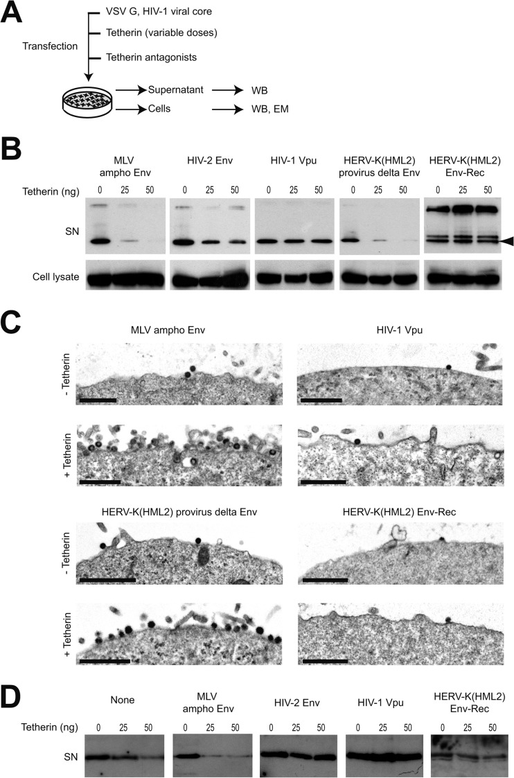
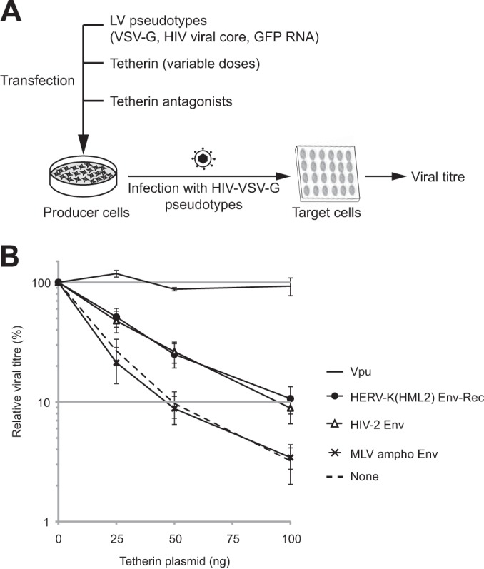
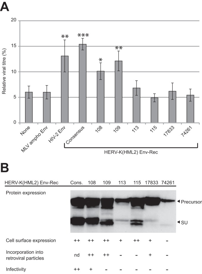
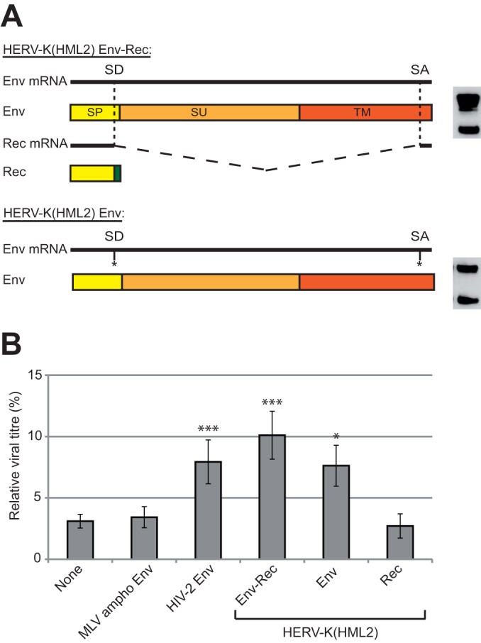
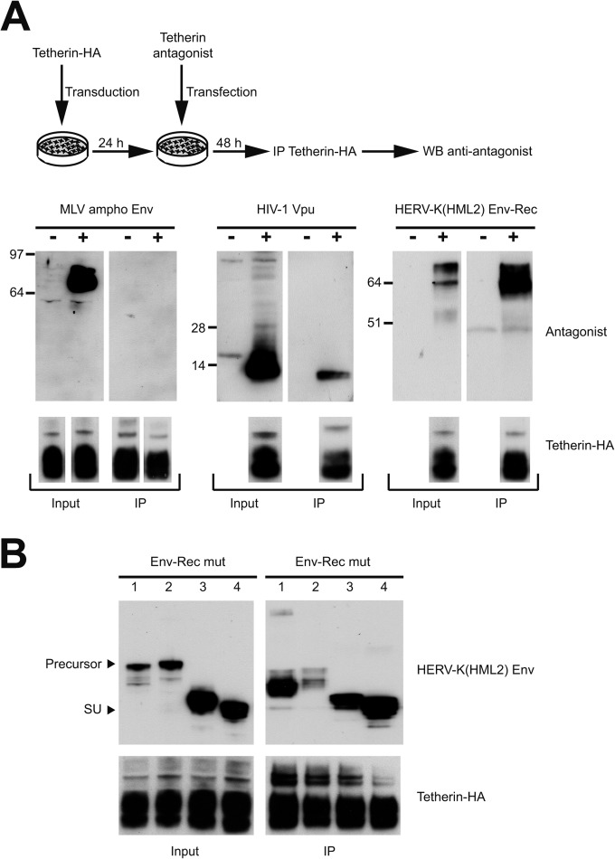
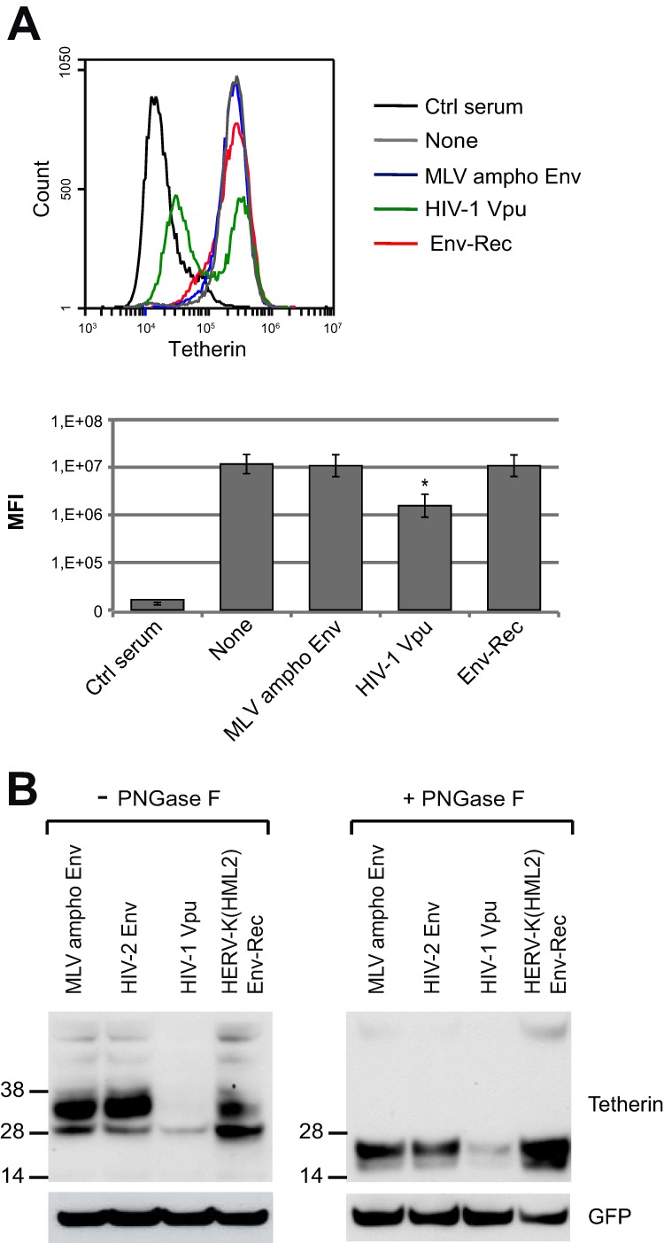
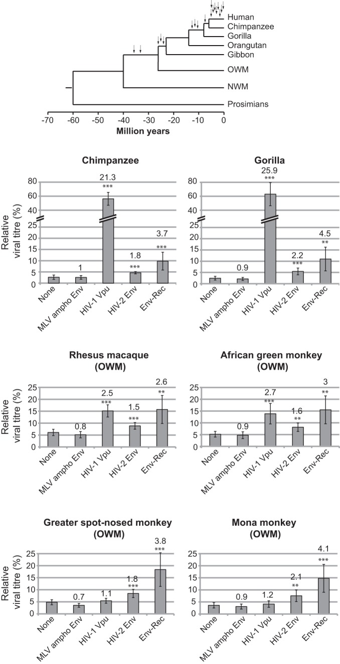
Endogenous retroviruses are the remnants of past retroviral infections that are scattered within mammalian genomes. In humans, most of these elements are old degenerate sequences that have lost their coding properties. The HERV-K(HML2) family is an exception: it recently amplified in the human genome and corresponds to the most active proviruses, with some intact open reading frames and the potential to encode viral particles. Here, using a reconstructed consensus element, we show that HERV-K(HML2) proviruses are able to inhibit Tetherin, a cellular restriction factor that is active against most enveloped viruses and acts by keeping the viral particles attached to the cell surface. More precisely, we identify the Envelope protein (Env) as the viral effector active against Tetherin. Through immunoprecipitation experiments, we show that the recognition of Tetherin is mediated by the surface subunit of Env. Similar to Ebola glycoprotein, HERV-K(HML2) Env does not mediate Tetherin degradation or cell surface removal; therefore, it uses a yet-undescribed mechanism to inactivate Tetherin. We also assessed all natural complete alleles of endogenous HERV-K(HML2) Env described to date for their ability to inhibit Tetherin and found that two of them (out of six) can block Tetherin restriction. However, due to their recent amplification, HERV-K(HML2) elements are extremely polymorphic in the human population, and it is likely that individuals will not all possess the same anti-Tetherin potential. Because of Tetherin's role as a restriction factor capable of inducing innate immune responses, this could have functional consequences for individual responses to infection.

Importance: Tetherin, a cellular protein initially characterized for its role against HIV-1, has been proven to counteract numerous enveloped viruses. It blocks the release of viral particles from producer cells, keeping them tethered to the cell surface. Several viruses have developed strategies to inhibit Tetherin activity, allowing them to efficiently infect and replicate in their host. Here, we show that human HERV-K(HML2) elements, the remnants of an ancient retroviral infection, possess an anti-Tetherin activity which is mediated by the envelope protein. It is likely that this activity was an important factor that contributed to the recent, human-specific amplification of this family of elements. Also, due to their recent amplification, HERV-K(HML2) elements are highly polymorphic in the human population. Since Tetherin is a mediator of innate immunity, interindividual variations among HERV-K(HML2) Env genes may result in differences in immune responses to infection.


Figures

FIG 1
FIG 1
Tetherin restriction is antagonized by HERV-K(HML2) Env-Rec. (A) Scheme of the experimental procedure. 293T cells were transfected with the indicated plasmids. Two days after transfection, cell supernatants were harvested and cells were either lysed to perform Western blot analysis or prepared for electron microscopy observation. (B) HIV-1 Gag protein content in neat supernatants (SN) was analyzed by Western blotting; p55 levels in corresponding cell lysates are displayed below for comparison. The nature of the potential Tetherin antagonist transfected is indicated above each gel part, and the doses of Tetherin transfected are given above each well (0, 25, or 50 ng). HIV-1 Gag p24 protein is indicated with an arrowhead. The additional bands observed in some supernatant samples (approximately 26 and 35 to 40 kDa) correspond to partially processed Gag protein. (C) Electron microscopy of cells obtained as described for panel A. Shown is a low magnification of cell membranes with viral particles observed by electron microscopy after negative staining. Images obtained with HIV-2 Env (not displayed here) were similar to those observed with HIV-1 Vpu and HERV-K(HML2) Env-Rec. Scale bars correspond to 1 μm. (D) HERV-K Gag protein content in neat supernatant was analyzed by Western blotting. The experimental procedure used was the same as that described for panel A, except the CMV-driven HERV-K(HML2) provirus defective for the envelope gene (instead of HIV-1 viral core) was transfected. Only cell supernatants were analyzed, since the anti-HERV-K(HML2) Gag antibody does not allow its specific detection in cell lysates (the signal/noise ratio is too high).
FIG 2
FIG 2
Quantitative assay of the HERV-K(HML2) Env-Rec-mediated inhibition of Tetherin activity. (A) Schematic description of the experimental procedure. 293T cells were transfected with the indicated plasmids. Three days posttransfection, the supernatants of producer cells were harvested to infect target cells. The percentage of infected cells was measured after 72 h by flow cytometry analysis, and viral titers were calculated for each condition. (B) The evolution of viral titers with the amount of transfected Tetherin (0 to 100 ng) is plotted for each potential inhibitor. The 100% value was set for each inhibitor using the titer measured without Tetherin. Error bars represent standard deviations of the means from 10 independent experiments, except for HIV-1 Vpu data (3 experiments).
FIG 3
FIG 3
Effect of endogenous alleles of HERV-K(HML2) Env-Rec on Tetherin activity. (A) Previously described endogenous alleles of HERV-K(HML2) Env-Rec were tested for their ability to antagonize Tetherin as described in the legend to Fig. 2A. For each protein tested, the relative viral titer measured with 100 ng of Tetherin is represented (100% corresponds to the value measured in the absence of Tetherin). Under these conditions, HIV-1 Vpu restores the viral titer to 95.3% of the no-Tetherin condition (not shown). For statistical tests, all results were compared to the “None” condition. Asterisks indicate values significantly different from that obtained under the “None” condition (unpaired two-tailed t test; *, P < 0.05; **, P < 0.01; ***, P < 0.001). (B) Recapitulation of the functional properties of the 6 endogenous alleles of HERV-K(HML2) Env-Rec. Expression levels of the different HERV-K(HML2) Env-Rec alleles were analyzed by Western blotting on 293T cell lysates following transient transfection. Other functional properties of these alleles, which have been previously assessed (19), also are recapitulated. Cons., consensus HERV-K(HML2) element; nd, not determined.
FIG 4
FIG 4
HERV-K(HML2) Env inhibits Tetherin activity. (A) The HERV-K(HML2) Env-Rec plasmid leads to the expression of 2 distinct proteins via alternative splicing: the full-length RNA encodes Env, a glycoprotein that is cleaved into 2 subunits during synthesis (surface subunit, SU, and transmembrane subunit, TM), while internal splicing sites (SD for splice donor and SA for splice acceptor) lead to the production of Rec, an accessory protein whose first exon (in yellow) is contained within the signal peptide (SP) of Env while the second exon (in green) is translated from a different reading frame. An expression vector leading to the production of Env but not Rec was generated by point mutations introduced in the splicing sites. Env expression levels obtained with each of the two constructs are shown on the right. (B) The ability of HERV-K(HML2) Env and Rec to antagonize Tetherin restriction was assayed as described in the legend to Fig. 2A. Results are given as described in the legend to Fig. 3A.
FIG 5
FIG 5
Characterization of HERV-K(HML2) Env domains necessary for inhibiting Tetherin activity. (A) Scheme of the different derivatives of HERV-K(HML2) Env-Rec. All modifications were designed to modify Env without altering the Rec ORF. The different domains of each construct are indicated: SP (signal peptide, yellow), SU (orange), and TM (red). HERV-K(HML2) Env-Rec mut1 corresponds to a C-terminal truncated version of the complete Env (41 aa deleted). HERV-K(HML2) Env-Rec mut2 has its membrane spanning (in green) and cytoplasmic domains replaced by a GPI anchor (the signal for the addition of the GPI anchor is depicted by a triangle in the scheme). HERV-K(HML2) Env-Rec mut3 consists of the SU subunit attached to the cell membrane by a GPI anchor. HERV-K(HML2) Env-Rec mut4 corresponds to the soluble surface subunit. For each construct, the expected protein structure is depicted on the right. (B) Expression levels of Env-Rec and derivatives were measured in transiently transfected 293T cells either by Western blotting on whole-cell lysates (left) or by FACS following cell surface staining (right). The histogram displays the median fluorescence intensities (MFI) of the whole population for each protein tested. (C) The efficiency of the different HERV-K(HML2) Env-Rec derivatives to antagonize Tetherin was assayed as described in the legend to Fig. 2A. The representation of results is the same as that described in the legend to Fig. 3A.
FIG 6
FIG 6
Assay for interaction between HERV-K(HML2) Env and Tetherin. (A) Scheme of the experimental procedure. 293T cells were transduced with an internally HA-tagged Tetherin lentiviral vector and then transfected with different Tetherin antagonists [MLV ampho Env, HIV-1 Vpu, and HERV-K(HML2) Env-Rec]. Cell lysates were harvested 48 h posttransfection and subjected to anti-HA immunoprecipitation. The presence of the Tetherin antagonists or control protein in the immunoprecipitated fraction (IP) was then assessed by Western blotting (top). A fraction of the cell lysates before immunoprecipitation (input) was included to ensure their efficient expression and detection. The presence of similar amounts of Tetherin in all samples (input and IP fractions) was checked by Western blotting in a separate gel run under the same conditions using an anti-HA antibody (lower). The minus antagonist sample was the same for each protein tested and was migrated only once to ensure Tetherin expression; it is presented below the MLV ampho panel. (B) In order to map the interaction domain between Tetherin and HERV-K(HML2) Env-Rec, HERV-K(HML2) Env-Rec derivatives (mut 1, mut 2, mut 3, and mut 4) were assayed for their ability to interact with Tetherin by following the protocol described for panel A. In the top panels, arrows point to the precursor form and SU subunit of HERV-K(HML2) Env-Rec derivatives. As described for panel A, Tetherin expression levels were measured for all samples by Western blotting and are presented below the Env panels.
FIG 7
FIG 7
Mechanistic aspects of HERV-K(HML2) Env-Rec inhibition of Tetherin. (A) Assay for cell membrane downregulation of Tetherin by inhibitors. HeLa cells were transfected with expression vectors for Tetherin inhibitors or control proteins. Forty-eight hours posttransfection, cells were stained for cell surface Tetherin. A staining with a control rabbit serum (Ctrl) also was included in the experiment. The histogram on the top corresponds to 1 representative experiment out of 3. Below is displayed the median fluorescence intensities for each antagonist tested. Error bars represent standard deviations of the means from three independent experiments. All results are compared to the “None” condition. Asterisks indicate values significantly different (*, P < 0.05) from that obtained under “None” conditions (unpaired two-tailed t test). (B) Assay for Tetherin degradation by inhibitors. 293T cells were transfected with 8.91, pHR′SIN-cPPT-SEW, VSV-G, Tetherin expression vector, and a Tetherin inhibitor or control protein. Two days after transfection, cell lysates were prepared, treated with PNGase F (right), or left untreated (left). The membrane was stained for Tetherin (top) and then stripped and stained again for GFP (expressed from transfected pHR′SIN-cPPT-SEW) (bottom) to ensure that each well of the gel had been equally loaded.
FIG 8
FIG 8
Effect of HERV-K(HML2) Env-Rec on several primate Tetherin proteins. HERV-K(HML2) Env-Rec was tested for its ability to antagonize several primate Tetherin proteins as described in the legend to Fig. 2A. For each Tetherin and antagonist tested, the relative viral titer measured with 25 ng of Tetherin is represented (100% corresponds to the value measured in the absence of Tetherin), with the average fold increase compared to the “None” condition indicated above each antagonist. Statistical tests were done as described in the legend to Fig. 3A using 9 independent experiments. A phylogenetic tree of primates is presented (top). The arrows indicate amplification bursts of HERV-K(HML2) elements during primate evolution (from reference 8) (OGM and NWM, Old and New World monkeys, respectively).

References

    1. Bannert N, Kurth R. 2004. Retroelements and the human genome: new perspectives on an old relation. Proc. Natl. Acad. Sci. U. S. A. 101(Suppl 2):S14572–S14579. 10.1073/pnas.0404838101. - DOI - PMC - PubMed
    1. Stoye JP. 2012. Studies of endogenous retroviruses reveal a continuing evolutionary saga. Nat. Rev. Microbiol. 10:395–406. 10.1038/nrmicro2783. - DOI - PubMed
    1. Feschotte C, Gilbert C. 2012. Endogenous viruses: insights into viral evolution and impact on host biology. Nat. Rev. Genet. 13:283–296. 10.1038/nrg3199. - DOI - PubMed
    1. Dewannieux M, Heidmann T. 2013. Endogenous retroviruses: acquisition, amplification and taming of genome invaders. Curr. Opin. Virol. 3:646–656. 10.1016/j.coviro.2013.08.005. - DOI - PubMed
    1. Blond JL, Lavillette D, Cheynet V, Bouton O, Oriol G, Chapel-Fernandes S, Mandrand B, Mallet F, Cosset FL. 2000. An envelope glycoprotein of the human endogenous retrovirus HERV-W is expressed in the human placenta and fuses cells expressing the type D mammalian retrovirus receptor. J. Virol. 74:3321–3329. 10.1128/JVI.74.7.3321-3329.2000. - DOI - PMC - PubMed

Publication types

MeSH terms