

Laser OptoAcoustic Tomography: Towards New Technology for Biomedical Diagnostics
- PMID: 25210212
- PMCID: PMC4157689
- DOI: 10.1016/j.nima.2012.12.035
Laser OptoAcoustic Tomography: Towards New Technology for Biomedical Diagnostics
Abstract
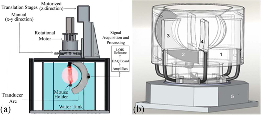
This paper provides a short review of physical principles, technology, biomedical applications and perspectives of the optoacoustic imaging. Ideas that made this rapidly developing field possible include the following: (1) laser pulses may be effectively used to produce acoustic pressure in biological tissues localized to the areas of increased optical absorption, (2) the resulting acoustic (ultrasonic) waves propagate in tissues with minimal distortions and attenuation, (3) 2D and 3D maps (images) of the absorbed optical energy can be reconstructed with high resolution from the detected optoacoustic signals. Modern optoacoustic imaging systems include scanning focused transducers and 2D/3D transducer arrays. The widely accepted 2D arrays are employed either for real-time 2D optoacoustic imaging or for 3D imaging via translational or rotational scanning. A commercial prototype of a 3D OAT system is being developed by TomoWave Labs where major biomedical applications include visualization of specific targeting using exogenous optoacoustic contrast agents and imaging of blood distribution and oxygentaion status can be investigated.
Keywords: gold nanorods; in-vivo imaging; optoacoustic tomography; preclinical research.
Figures
Similar articles
-
Melanin-Based Contrast Agents for Biomedical Optoacoustic Imaging and Theranostic Applications.Int J Mol Sci. 2017 Aug 7;18(8):1719. doi: 10.3390/ijms18081719. Int J Mol Sci. 2017. PMID: 28783106 Free PMC article. Review.
-
Stochastic three-dimensional numerical phantoms to enable computational studies in quantitative optoacoustic computed tomography of breast cancer.J Biomed Opt. 2023 Jun;28(6):066002. doi: 10.1117/1.JBO.28.6.066002. Epub 2023 Jun 20. J Biomed Opt. 2023. PMID: 37347003 Free PMC article.
-
Focused array transducer for two-dimensional optoacoustic tomography.J Acoust Soc Am. 2004 Sep;116(3):1498-506. doi: 10.1121/1.1781710. J Acoust Soc Am. 2004. PMID: 15478415
-
Melanin nanoparticles as a novel contrast agent for optoacoustic tomography.Photoacoustics. 2015 Feb 14;3(1):35-43. doi: 10.1016/j.pacs.2015.02.001. eCollection 2015 Mar. Photoacoustics. 2015. PMID: 25893172 Free PMC article.
-
Emerging contrast agents for multispectral optoacoustic imaging and their biomedical applications.Chem Soc Rev. 2021 Jul 21;50(14):7924-7940. doi: 10.1039/d1cs00358e. Epub 2021 Jun 11. Chem Soc Rev. 2021. PMID: 34114588 Review.
Cited by
-
Imaging tumor acidosis: a survey of the available techniques for mapping in vivo tumor pH.Cancer Metastasis Rev. 2019 Jun;38(1-2):25-49. doi: 10.1007/s10555-019-09782-9. Cancer Metastasis Rev. 2019. PMID: 30762162 Free PMC article. Review.
-
Role of blood oxygenation saturation in ovarian cancer diagnosis using multi-spectral photoacoustic tomography.J Biophotonics. 2021 Apr;14(4):e202000368. doi: 10.1002/jbio.202000368. Epub 2021 Jan 6. J Biophotonics. 2021. PMID: 33377620 Free PMC article.
-
Photostability of Contrast Agents for Photoacoustics: The Case of Gold Nanorods.Nanomaterials (Basel). 2021 Jan 6;11(1):116. doi: 10.3390/nano11010116. Nanomaterials (Basel). 2021. PMID: 33419130 Free PMC article. Review.
-
Intrinsic beam emittance of laser-accelerated electrons measured by x-ray spectroscopic imaging.Sci Rep. 2016 Apr 19;6:24622. doi: 10.1038/srep24622. Sci Rep. 2016. PMID: 27090440 Free PMC article.
-
Review of cost reduction methods in photoacoustic computed tomography.Photoacoustics. 2019 Jul 26;15:100137. doi: 10.1016/j.pacs.2019.100137. eCollection 2019 Sep. Photoacoustics. 2019. PMID: 31428558 Free PMC article. Review.
References
-
- Oraevsky AA, Karabutov AA. Optoacoustic Tomography. In: Vo-Dinh T, editor. Biomedical Photonics Handbook. Boca Raton: CRC Press; 2003. pp. 34/21–34/34.
-
- Ermilov SA, Khamapirad T, Conjusteau A, Leonard MH, Lacewell R, Mehta K, Miller T, Oraevsky AA. Laser optoacoustic imaging system for detection of breast cancer. Journal of biomedical optics. 2009;14:024007. - PubMed
Grants and funding
LinkOut - more resources
Full Text Sources
Other Literature Sources