

Role of 3D-CT for orthodontic and ENT evaluation in Goldenhar syndrome
- PMID: 25210224
- PMCID: PMC4157534
Role of 3D-CT for orthodontic and ENT evaluation in Goldenhar syndrome
Abstract
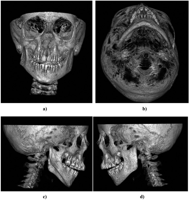
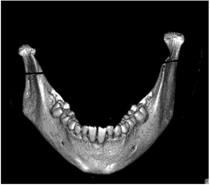
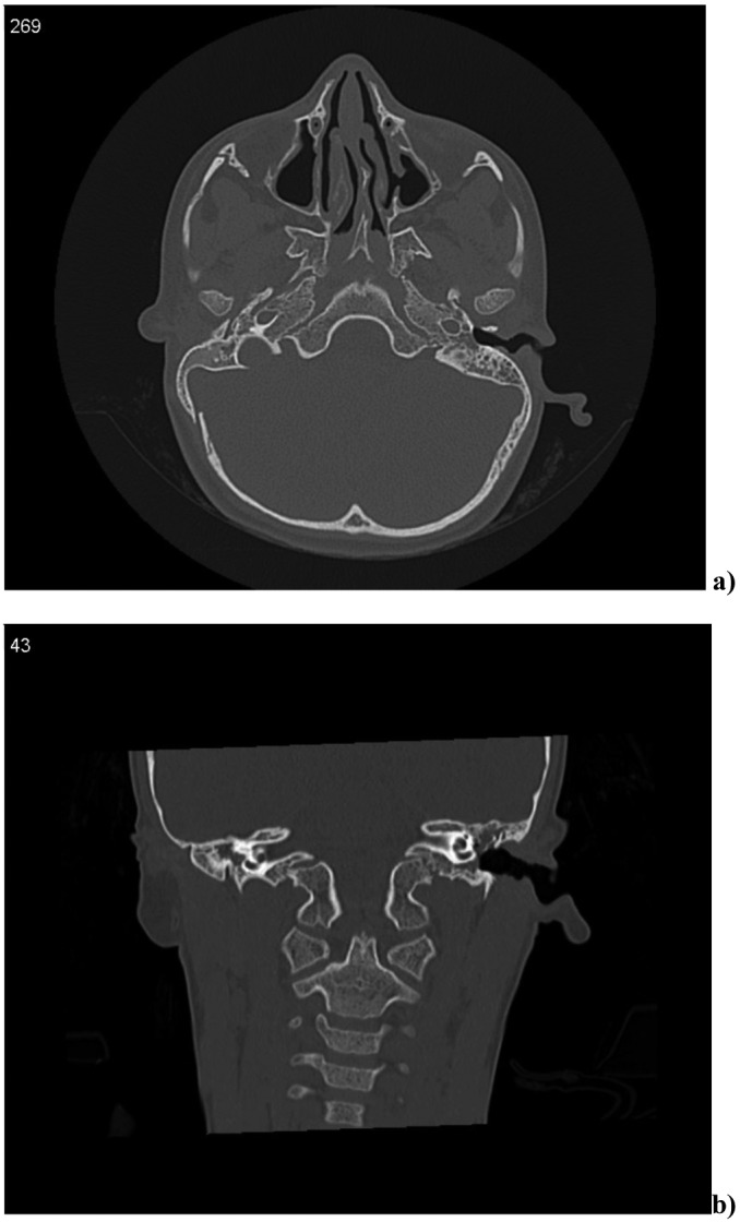
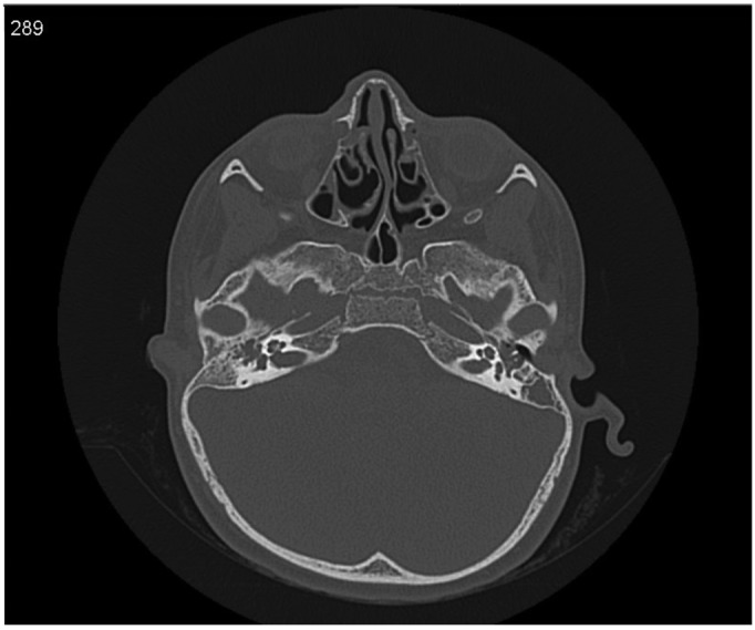
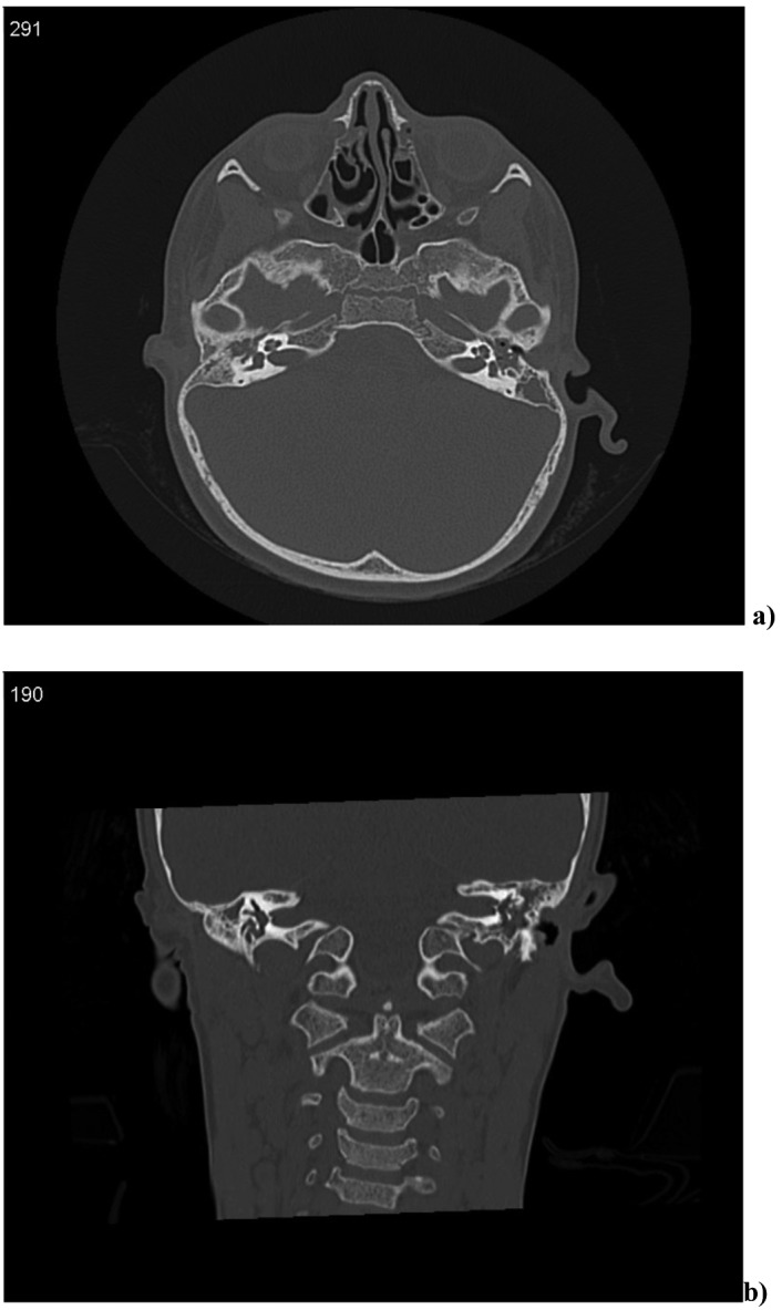
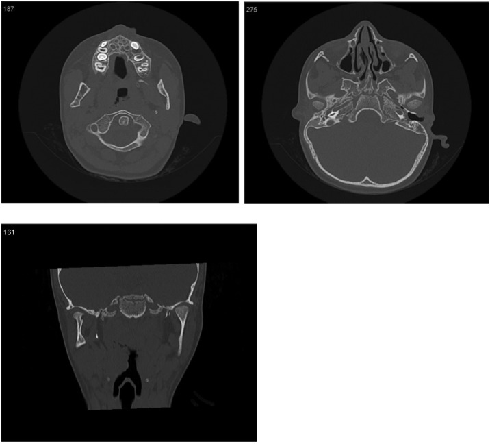
Goldenhar syndrome is a congenital condition that includes anomalies of the derivatives of the first and second brachial arches, vertebral defects and ocular abnormalities. It is also known as oculo-auriculo-vertebrale syndrome (OAVS), hemifacial microsomia, or first or second brachial arch syndrome. It was first described by Van Duyse in 1882 and better studied by M. Goldenhar in 1952. Its treatment requires a multidisciplinary approach. Herein, we describe the value of 3D-CT evaluation in a patient with Goldenhar syndrome, with particular regard to planning diagnostic and therapeutic approach. A 7-year-old boy with Goldenhar syndrome with definite post-natal genetic diagnosis was referred to our Department of Radiology for neuroimaging of the temporal bone. By 3D-CT evaluation of this young patient we observed the asymmetry of the condyles with the right one dysmorphic, short and wide; the auricle of the right ear was replaced by a dysmorphic rough; the right middle ear had a hypoplastic tympanic cavity and the internal auditory canal of right ear was atresic. In our experience, 3D-CT is a powerful diagnostic instrument and offers many advantages: volumetric reproduction of cranium and soft tissues, no overlap of anatomic parts that limits the visibility of various structures, high precision and assurance of images, and a constant and easily reproducible reference system. In our case, 3D-CT offered a very complete evaluation of all malformations of mandibular and temporal bone that characterize this syndrome and representing an important step for ENT and orthodontic therapeutic approaches.
La sindrome di Goldenhar è una condizione congenita associata a difetti di sviluppo del primo e secondo arco branchiale, anomalie oculari e vertebrali. Possono essere associate anche anomalie cardiache, renali e del sistema nervoso centrale. Anche conosciuta come Sindrome oculo-auriculo-vertebrale, microsomia emifacciale o sindrome del primo e secondo arco branchiale. Fu descritta per la prima volta da Van Duyse nel 1882 e successivamente studiato da M. Goldenhar nel 1952 in modo più dettagliato. Il suo trattamento richiede un approccio multidisciplinare. Nel presente articolo si descrive il valore della TC-3D in un giovane paziente con sindrome di Goldenhar, con particolare riferimento alla diagnosi e alla pianificazione dell'approccio terapeutico. Un paziente di 7 anni con diagnosi postnatale geneticamente confermata di sindrome di Goldenhar è stato sottoposto a TC-3D presso il dipartimento di radiologia del nostro Policlinico. La valutazione radiologica ha consentito di mettere in evidenza l'asimmetria dei condili mandibolari (il destro appariva dismorfico, corto e largo), il padiglione dell'orecchio omolaterale sostituito da un rudimento dismorfico, l'orecchio medio di destra presentava una cavità timpanica ipoplasica con condotto uditivo interno atresico. Secondo la nostra esperienza la TC-3D rappresenta un potente strumento di diagnosi e offre molti vantaggi consentendo una precisa rappresentazione volumetrica dei tessuti del cranio e dei tessuti molli senza sovrapposizione di parti anatomiche garantendo un'elevata precisione delle immagini. Nel nostro caso la TC-3D offre una rappresentazione completa di tutte le malformazioni sia mandibolari che dell'osso temporale che caratterizzano la Sindrome di Gholdenar presupposto fondamentale per la pianificazione dell'approccio terapeutico ortodontico e otorinolaringoiatrico.
Keywords: 3D-CT; Facial asymmetry; Goldenhar Syndrome.
Figures

References
-
- Kokavec R. Goldenhar syndrome with various clinical manifestations. Cleft Palate Craniofac J. 2006;43:628–634. - PubMed
-
- Goldenhar M. Malformative associations of eye and ear, expecially the dermoid syndrome and the epibulbar dermoidauricular appendix-preauricula fistula auris congenital syndrome and its connections with mandibular-facial-dysostosis. J Genet Hum. 1952;1:243–282.
-
- Garcia de Paula e Silva FW, Carvalho FK, Diaz-Serrano KV, et al. Solitary median maxillary central incisor in association with Goldenhar's syndrome: a case report. Spec Care Dentist. 2007;27:105–107. - PubMed
-
- Vinay C, Reddy RS, Uloopi KS, et al. Craniofacial features in Goldenhar syndrome. J Indian Soc Pedod Prev Dent. 2009;27:121–124. - PubMed
-
- Bielicka B. Interdisciplinary treatment of patients with Goldenhar syndrome - clinical reports. Dent Med Probl. 2006;43:458–462.
Publication types
MeSH terms
LinkOut - more resources
Full Text Sources
Medical
Research Materials
Miscellaneous