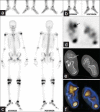
Incremental value of single photon emission tomography/computed tomography in 3-phase bone scintigraphy of an accessory navicular bone
- PMID: 25210293
- PMCID: PMC4157201
- DOI: 10.4103/0972-3919.136600
Incremental value of single photon emission tomography/computed tomography in 3-phase bone scintigraphy of an accessory navicular bone
Abstract
Accessory navicular bone is one of the supernumerary ossicles in the foot. Radiography is non diagnostic in symptomatic cases. Accessory navicular has been reported as a cause of foot pain and is usually associated with flat foot. Increased radio tracer uptake on bone scan is found to be more sensitive. We report a case highlighting the significance of single photon emission tomography/computed tomography in methylene diphosphonate bone scan in the evaluation of symptomatic accessory navicular bone where three phase bone scan is equivocal.
Keywords: 99mTc-methylene diphosphonate; Accessory navicular bone; bone scan; single photon emission tomography/computed tomography.
Conflict of interest statement
Figures

References
-
- Groshar D, Gorenberg M, Ben-Haim S, Jerusalmi J, Liberson A. Lower extremity scintigraphy: The foot and ankle. Semin Nucl Med. 1998;28:62–77. - PubMed
-
- Romanowski CA, Barrington NA. The accessory navicular: An important cause of medial foot pain. Clin Radiol. 1992;46:261–4. - PubMed
-
- Fredrick LA, Beall DP, Ly JQ, Fish JR. The symptomatic accessory navicular bone: A report and discussion of the clinical presentation. Curr Probl Diagn Radiol. 2005;34:47–50. - PubMed
-
- Chuang YW, Tsai WS, Chen KH, Hsu HC. Clinical use of high-resolution ultrasonography for the diagnosis of type II accessory navicular bone. Am J Phys Med Rehabil. 2012;91:177–81. - PubMed
LinkOut - more resources
Full Text Sources
Other Literature Sources

