C1-2 posterior arthrodesis technique with a left segmental and right transarticular fixation. A hybrid novel (Kotil) technique
- PMID: 25210344
- PMCID: PMC4158630
- DOI: 10.4103/0974-8237.139213
C1-2 posterior arthrodesis technique with a left segmental and right transarticular fixation. A hybrid novel (Kotil) technique
Abstract
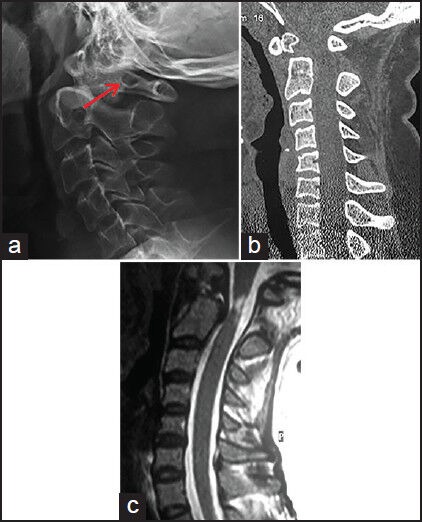
Introduction: The most commonly used techniques for C1-C2 posterior arthrodesis are Goel and Magerl fixation techniques. Due to the anatomical variations of the region, the prior determination of the surgical technique might be hard. Right side Magerl, left side Goel's C1-C2 posterior arthrodesis case is presented as a new surgical combination technique used due to anatomical difficulties.
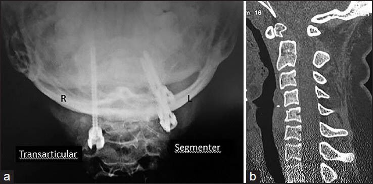
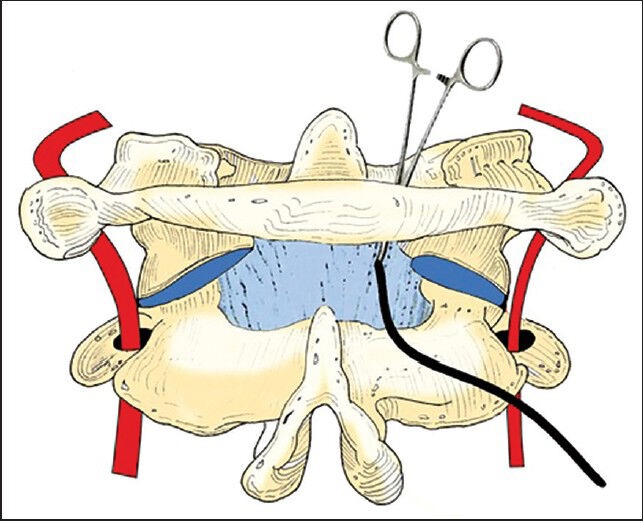
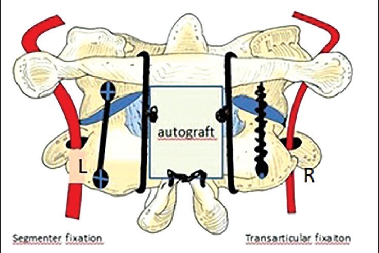
Materials and methods: Posterior C1-C2 arthrodesis operation was indicated for a 56-year-old female patient for the treatment of atlanto-axial subluxation caused by os odontoideum. First it was fixed from the nondominant arterial side (right vertebral artery) with Magerl (transarticular) technique. The left side was not suitable for the anatomical transarticular fixation, and the contralateral Goel fixation technique (segmental) was performed. Eventually, right side transarticular left side segmental fixation techniques were combined in one patient for the first time and C1-C2 fusion combination technique was presented.
Results: Both Goel and Magerl techniques of C1-C2 posterior fusion techniques were successfully used simultaneously. The operation was initiated with Magerl technique with one screw on the nondominant side. The contralateral side was not suitable for Magerl technique therefore we changed to Goel's technique. Although, fluoroscopy was used 3 times as much during the introduction of the Drill with Magerl technique, twice as much operative time was spent during hemostasis and bleeding, preparation of the C1 entry point, and the reconstruction of polyaxial screws for Goel technique. No neurovascular complications were occurred during both procedures.
Discussion: Combination of two C1-C2 posterior fusion techniques, Goel and Magerl, in suitable cases caused by anatomical or other reasons appears to be an alternative surgical procedure that protects the patient from complications. For a collection of better data, other studies that include large numbers of patients with high evidential value should be conducted.
Keywords: C1-C2 posterior arthrodesis; Kotil technique; novel technique; segmental fixation; transarticular fixation.
Conflict of interest statement
Figures
Similar articles
-
[Harms technique of C1-C2 fixation with polyaxial screws and rods].Acta Chir Orthop Traumatol Cech. 2005;72(1):22-7. Acta Chir Orthop Traumatol Cech. 2005. PMID: 15860148 Czech.
-
Clinically relevant complications related to posterior atlanto-axial fixation in atlanto-axial instability and their management.Clin Neurol Neurosurg. 2014 Aug;123:131-5. doi: 10.1016/j.clineuro.2014.05.020. Epub 2014 Jun 4. Clin Neurol Neurosurg. 2014. PMID: 25012025
-
New technique for C1-C2 fixation.Surg Neurol Int. 2018 May 7;9:94. doi: 10.4103/sni.sni_10_18. eCollection 2018. Surg Neurol Int. 2018. PMID: 29888028 Free PMC article.
-
A biomechanical study of unilateral posterior atlantoaxial transarticular screw fixation.J Long Term Eff Med Implants. 2005;15(1):33-8. doi: 10.1615/jlongtermeffmedimplants.v15.i1.40. J Long Term Eff Med Implants. 2005. PMID: 15715514
-
Outcome comparison of atlantoaxial fusion with transarticular screws and screw-rod constructs: meta-analysis and review of literature.J Spinal Disord Tech. 2014 Feb;27(1):11-28. doi: 10.1097/BSD.0b013e318277da19. J Spinal Disord Tech. 2014. PMID: 23128387 Review.
References
-
- Papagelopoulos PJ, Currier BL, Hokari Y, Neale PG, Zhao C, Berglund LJ, et al. Biomechanical comparison of C1-C2 posterior arthrodesis techniques. Spine (Phila Pa 1976) 2007;32:E363–70. - PubMed
-
- Naderi S, Crawford NR, Song GS, Sonntag VK, Dickman CA. Biomechanical comparison of C1-C2 posterior fixations. Cable, graft, and screw combinations. Spine (Phila Pa 1976) 1998;23:1946–55. - PubMed
-
- Goel A, Laheri V. Plate and screw fixation for atlanto-axial subluxation. Acta Neurochir (Wien) 1994;129:47–53. - PubMed
-
- Goel A, Desai K, Muzumdar D. Atlantoaxial fixation using plate and screw method. A report of 160 treated patients. Neurosurgery. 2002;51:1351–7. - PubMed
-
- Magerl F, Seemann PS. Stable posterior fusion of the atlas and axis by transarticular screw fixation. In: Kehr P, Weidner A, editors. Cervical Spine I. New York: Springer Wien; 1986. pp. 322–7.
Publication types
LinkOut - more resources
Full Text Sources
Other Literature Sources
Miscellaneous