. 2014 Aug 26:8:1611-21.
doi: 10.2147/OPTH.S60893. eCollection 2014.

Clinical utilization of anti-vascular endothelial growth-factor agents and patient monitoring in retinal vein occlusion and diabetic macular edema

Affiliations

Clinical utilization of anti-vascular endothelial growth-factor agents and patient monitoring in retinal vein occlusion and diabetic macular edema

Szilárd Kiss et al. Clin Ophthalmol. .

Abstract

Purpose: To examine the utilization of bevacizumab and ranibizumab and disease monitoring in patients with branch or central retinal vein occlusion (BRVO/CRVO) or diabetic macular edema (DME) in clinical practice.

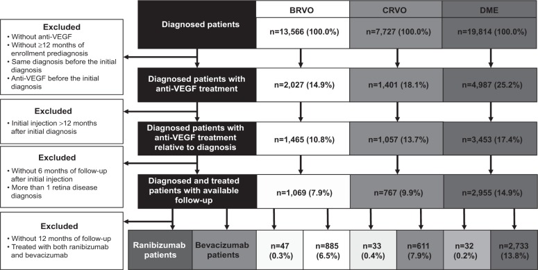
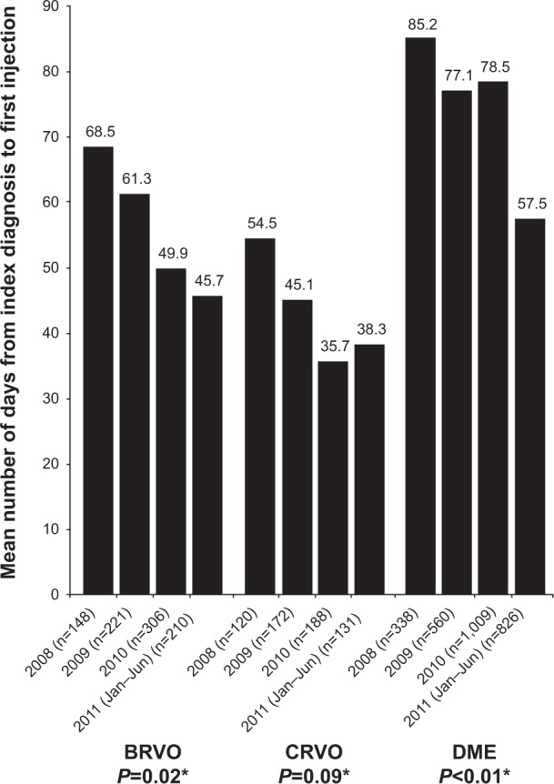
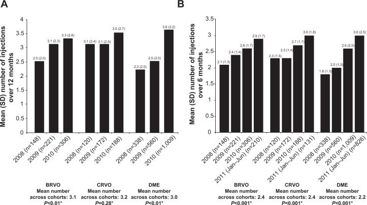
Patients and methods: This retrospective claims analysis included newly diagnosed patients with one or more bevacizumab or ranibizumab injections. Bevacizumab or ranibizumab utilization was assessed by year of first injection: 2008-2010 cohorts (12-month follow-up), January to June 2011 cohort (6-month follow-up). The main outcome measures were mean annual numbers of injections, ophthalmologist visits and optical coherence tomography examinations, and proportion of patients with additional laser or intravitreal triamcinolone (IVTA) use.

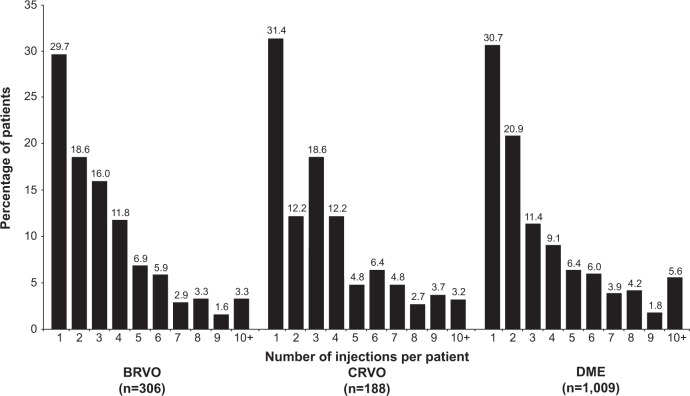
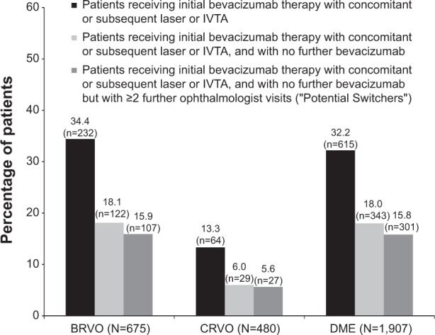
Results: A total of 885 BRVO, 611 CRVO, and 2,733 DME patients treated with bevacizumab were included, with too few ranibizumab-treated patients for meaningful analysis. Across the 2008, 2009, and 2010 cohorts, mean annual numbers of bevacizumab injections increased, but remained low (BRVO 2.5, 3.1, 3.3; CRVO 3.1, 3.1, 3.5; and DME 2.2, 2.5, 3.6, respectively); mean ophthalmologist visits ranged between 4.4 and 6.5, and mean optical coherence tomography examinations ranged between 3.1 and 3.9 across all conditions. A total of 42.0% of BRVO, 16.5% of CRVO, and 57.7% of DME patients received additional laser or IVTA therapy. The number of bevacizumab injections was positively associated with laser use in BRVO (3.3 versus 2.9, P<0.03), and with laser or IVTA use in DME (laser, 3.3 versus 2.7, P<0.03; IVTA, 3.3 versus 3.0, P<0.05).

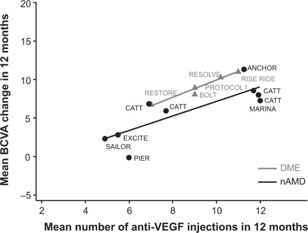
Conclusion: During the study period (2008-2011), bevacizumab was the main anti-VEGF therapy used in clinical practice for BRVO, CRVO, and DME. Patients treated with bevacizumab were monitored less frequently and received fewer injections than patients in major clinical trials of ranibizumab.

Keywords: anti-vascular endothelial growth factor; bevacizumab; diabetic macular edema; intravitreal; ranibizumab; retinal vein occlusion.


Figures

Figure 1
Figure 1
Patient flowchart. Notes: Of the 13,566 BRVO-, 7,727 CRVO-, and 19,814 DME-diagnosed patients, 932, 644, and 2,765, respectively, met all inclusion and exclusion criteria, with most patients receiving bevacizumab. The low number of ranibizumab users in each group (47, 33, and 32, respectively) precluded a meaningful analysis for this agent. Abbreviations: BRVO, branch retinal vein occlusion; CRVO, central retinal vein occlusion; DME, diabetic macular edema.
Figure 2
Figure 2
Mean days from index diagnosis to first bevacizumab injection, by diagnosis group and cohort. Notes: *One-way analysis of variance comparing mean time across cohorts. Among those with a diagnosis of BRVO, there was a significant decrease in the mean days from initial diagnosis to first injection: 68.5 days in 2008, 61.3 days in 2009, 49.9 days in 2010, and 45.7 days in 2011 (January–June cohort, P=0.02). Although a decrease in time from initial diagnosis to first injection was observed in those with a diagnosis of CRVO (mean 54.5 days in 2008, 45.1 days in 2009, 35.7 days in 2010, and 38.3 days in 2011 [January–June cohort]), the differences in mean time across the cohorts were not significant (P=0.09). In the DME group, the mean time from initial diagnosis to first injections decreased significantly over time (from mean of 85.2 days in 2008, to 77.1 days in 2009, 78.5 days in 2010, and 57.5 days in 2011 [January–June cohort]; P<0.01). Abbreviations: BRVO, branch retinal vein occlusion; CRVO, central retinal vein occlusion; DME, diabetic macular edema.
Figure 3
Figure 3
(A) Mean number (standard deviation [SD]) of bevacizumab injections over 12 months, by diagnosis group and cohort; (B) mean number (SD) of bevacizumab injections over 6 months, by diagnosis group and cohort. Notes: *One-way analysis of variance comparing mean time across cohorts. In each diagnosis group, the mean number of injections administered over the 12-month period after the first injection (index date) increased, with mean differences across the cohorts being statistically significant in the BRVO and DME groups, but not in the CRVO group. Abbreviations: BRVO, branch retinal vein occlusion; CRVO, central retinal vein occlusion; DME, diabetic macular edema.
Figure 4
Figure 4
Distribution of the number of injections over 12 months in the 2010 cohort, by diagnosis group. Notes: In analyses of the distribution of the number of injections in the 2010 cohort, small percentages of patients in each diagnosis group received ≤10 injections during the 12 months after their index diagnosis. Abbreviations: BRVO, branch retinal vein occlusion; CRVO, central retinal vein occlusion; DME, diabetic macular edema.
Figure 5
Figure 5
Use of laser and/or intravitreal triamcinolone relative to the timing of bevacizumab use during the first 12 months following the index date, by diagnosis group (2008–2010 cohorts). Notes: In those with BRVO, CRVO, and DME, approximately 15.9%, 5.6%, and 15.8%, respectively, of the total diagnosis group were considered to have switched to either laser or IVTA treatment from bevacizumab treatment (ie, received laser or IVTA at the time of or after bevacizumab treatment, and then discontinued bevacizumab but continued to visit an ophthalmologist for at least two visits). Abbreviations: BRVO, branch retinal vein occlusion; CRVO, central retinal vein occlusion; DME, diabetic macular edema; IVTA, intravitreal triamcinolone.
Figure 6
Figure 6
Correlations between number of bevacizumab and ranibizumab injections and mean ETDRS letters gained in 12 months in major published Phase II and III clinical trials in nAMD and DME. Note: Evidence from major prospective clinical trials suggests a positive correlation between the administration frequency of bevacizumab or ranibizumab and visual improvement. Abbreviations: BCVA, best-corrected visual acuity; DME, diabetic macular edema; ETDRS, Early Treatment Diabetic Retinopathy Study; nAMD, neovascular age-related macular degeneration.

References

    1. Romero-Aroca P. Managing diabetic macular edema: the leading cause of diabetes blindness. World J Diabetes. 2011;2(6):98–104. - PMC - PubMed
    1. Yau JW, Lee P, Wong TY, Best J, Jenkins A. Retinal vein occlusion: an approach to diagnosis, systemic risk factors and management. Intern Med J. 2008;38(12):904–910. - PubMed
    1. Turello M, Pasca S, Daminato R, et al. Retinal vein occlusion: evaluation of “classic” and “emerging” risk factors and treatment. J Thromb Thrombolysis. 2010;29(4):459–464. - PubMed
    1. Institute for Clinical and Economic Review (ICER) Technology Assessment Report: Anti-Vascular Endothelial Growth Factor Treatment for Diabetic Macular Edema. Boston: ICER; 2012. [Accessed June 10, 2013]. Available from: http://www.cms.gov/Medicare/Coverage/DeterminationProcess/downloads/id85....
    1. Rogers S, McIntosh RL, Cheung N, et al. The prevalence of retinal vein occlusion: pooled data from population studies from the United States, Europe, Asia, and Australia. Ophthalmology. 2010;117(2):313–319. - PMC - PubMed

LinkOut - more resources