

Case Reports
doi: 10.2147/OPTH.S67855.
eCollection 2014.
Endophthalmitis following pars plana vitrectomy for vitreous floaters
Affiliations
- PMID: 25210434
- PMCID: PMC4155899
- DOI: 10.2147/OPTH.S67855
Item in Clipboard
Case Reports
Endophthalmitis following pars plana vitrectomy for vitreous floaters
Clin Ophthalmol.
.
Abstract
A case of Staphylococcus caprae endophthalmitis in a young patient following pars plana vitrectomy for symptomatic vitreous floaters is reported here. Recent literature suggests that there is an increasing trend of performing pars plana vitrectomy for symptomatic floaters. Although rare, the potential risk of endophthalmitis should be explicitly discussed with patients considering surgical intervention for vitreous floaters.
Keywords: endophthalmitis; floaterectomy; pars plana vitrectomy; posterior vitreous detachment; vitreous floaters.
Figures

Postoperative day 1 appearance. Notes: (A) Fundus photography, right eye, demonstrating mildly hazy media with vascular sheathing and perivascular hemorrhages. (B) Spectral domain optical coherence tomography, right eye, demonstrating retinal thickening and irregular intraretinal precipitates. (C) Early-phase fluorescein angiography, right eye, demonstrating areas of blockage corresponding to areas of perivascular hemorrhage. (D) Late phase fluorescein angiography, right eye, revealing leakage from retinal vessels and from the optic disc.
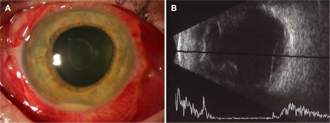
Postoperative day 4 appearance. Notes: (A) Anterior segment photography, right eye, demonstrating improving hypopyon and retracting fibrin clot. (B) B-scan echography, right eye, demonstrating low-lying choroidal detachment.

Postoperative month 9 appearance. Notes: (A) Fundus photography, right eye, demonstrating clear media and mottling of the retinal pigment epithelium. (B) Spectral domain optical coherence tomography, right eye, demonstrating an irregular retinal surface but intact ellipsoid layer.
References
-
- Park JC, Ramasamy B, Shaw S, Prasad S, Ling RH. A prospective and nationwide study investigating endophthalmitis following pars plana vitrectomy: incidence and risk factors. Br J Ophthalmol. 2014;98(4):529–533. - PubMed
-
- Weber-Krause B, Eckardt C. Incidence of posterior vitreous detachment in the elderly. Ophthalmologe. 1997;94(9):619–623. German. - PubMed
-
- Fijalkowsi N, Pershing S, Moshfeghi DM. The importance of keeping a broad differential in retina clinic: the spectrum of ophthalmic disease seen by retina specialists in a tertiary outpatient clinic setting. Ophthalmic Surg Lasers Imaging Retina. 2013;44(2):133–139. - PubMed
-
- Wagle AM, Lim WY, Yap TP, Neelam K, Au Eong HG. Utility values associated with vitreous floaters. Am J Ophthalmol. 2011;152(1):60–65. - PubMed
Publication types
Grants and funding
LinkOut - more resources
Full Text Sources
Other Literature Sources
Miscellaneous
