. 2014 Sep 6;10(9):1007-17.
doi: 10.7150/ijbs.9997. eCollection 2014.

The prostate basal cell (BC) heterogeneity and the p63-positive BC differentiation spectrum in mice

Affiliations

The prostate basal cell (BC) heterogeneity and the p63-positive BC differentiation spectrum in mice

Dong-Kee Lee et al. Int J Biol Sci. .

Abstract

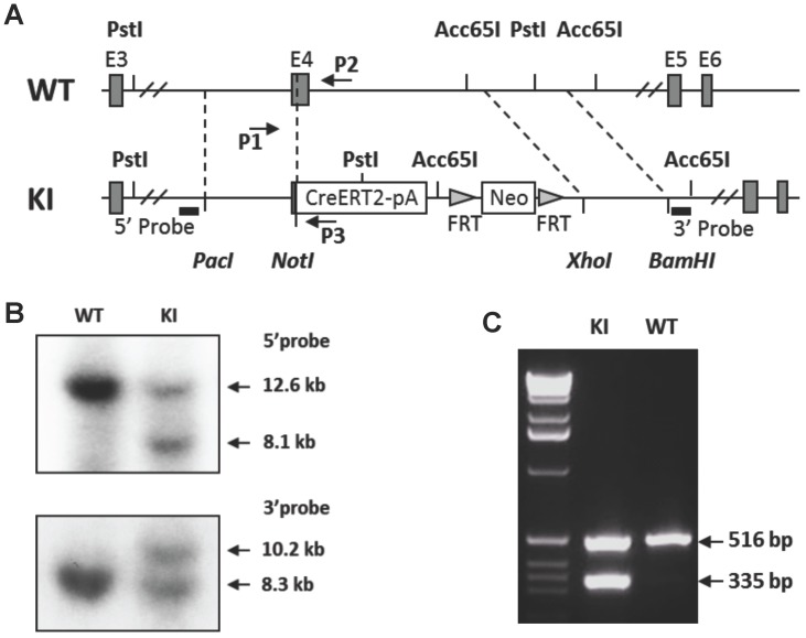
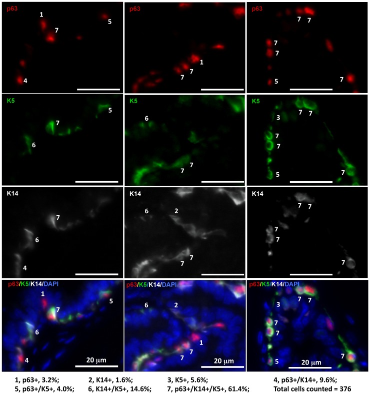
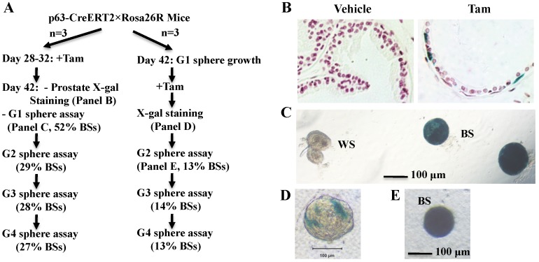
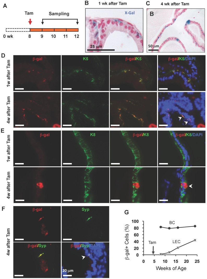
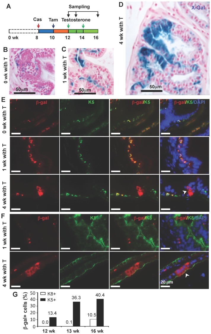
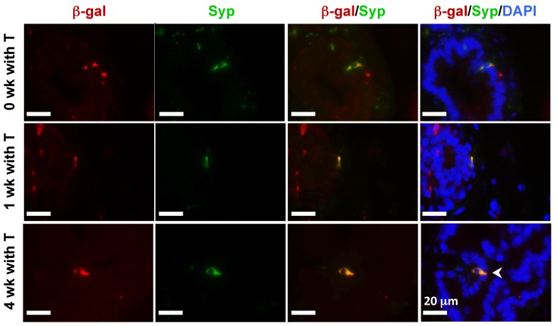
The prostate epithelium is composed of basal (BC), luminal (LEC), and neuroendocrine (NEC) cells. It is unclear how many subtypes of BCs in the prostate and which subtype of BCs contains the main stem cell niche in the adult prostate. Here we report seven BC subpopulations according to their p63, cytokeratin 14 (K14) and K5 expression patterns, including p63-positive/K14-negative/K5-negative (p63+/K14-/K5-), p63-/K14+/K5-, p63-/K14-/K5+, p63+/K14+/K5-, p63+/K14-/K5+, p63-/K14+/K5+, and p63+/K14+/K5+ BCs. We generated a p63-CreERT2 knock-in mouse line that expresses tamoxifen-inducible Cre activity in the p63-expressing cells, including the prostate BCs. We then crossbred this line with ROSA26R mice, and generated p63-CreERT2×ROSA26R bi-genic mice harboring the Cre-activated β-galactosidase reporter gene. We treated these bi-genic mice with tamoxifen to mark the p63+ BCs at different ages or under different hormonal conditions, and then traced the lineage differentiation of these genetically labeled BCs. We discovered that these p63+ BCs contain self-renewable stem cells in culture and efficiently differentiated into LECs, NECs and BCs in the postnatal, adult and re-generating mouse prostates. Therefore, BC population contains heterogeneous BCs that express different combinations of the p63, K14 and K5 differentiation markers. Because K14+ and K5+ BCs were previously shown to be extremely inefficient to produce LECs in adulthood, we propose that the p63+/K5-/K14- subpopulation of BCs contains most stem-like cells, especially in adult animals.

Keywords: Prostate epithelium; basal cell differentiation; lineage tracing; p63; stem cell..


Conflict of interest statement

Competing Interests: The authors have declared that no competing interest exists.

Figures

Figure 1
Figure 1
Generation of the p63-CreERT2 knock-in mouse line. A. Schematic drawing of the murine p63 gene wild type (WT) allele and the CreERT2-pA-FRT-Neo-FRT knock-in (KI) allele. Exons (E) 3-6 are indicated in both alleles. The 5' and 3' homologous regions used in the targeting vector are indicated by dotted lines between WT and KI alleles. The locations of the 5' and 3' probes used in Southern blot are indicated. The locations of primers P1, P2 and P3 used for genotype analysis are also indicated. B. Southern blot analysis of the WT and KI alleles. The 5' probe-detected 12.6 and 8.1 kb fragments were respectively derived from the WT and KI alleles after Pst I digestion. The 3' probe-detected 10.2 and 8.3 kb fragments were respectively derived from the KI and WT alleles after Acc65 I digestion. C. Genotype analysis of mouse genotypes by PCR. Mouse genomic DNA was used as PCR template. The 516 bp fragment was detected from the WT allele by primers P1 and P2. The 335 bp fragment was detected from the KI allele by primers P1 and P3.
Figure 2
Figure 2
Identification of seven subtypes of BCs in the adult mouse prostate by triple immunofluorescent staining of p63, K5 and K14. Triple immunofluorescent staining was performed on prostate sections prepared from three 6-week-old mice. Immunofluorescent staining signals of p63, K5 and K14 were separately imaged by using appropriate filters and a monochrome CCD camera and respectively labeled as red, green and white colors. Each of the three bottom panels is the merged image of the three panels above plus the blue DAPI staining signal for nuclei. The 7 BC subtypes were distinguished according to the expression patterns of p63, K5 and K14 as indicated. The percentage of each subtype BCs was calculated by normalizing to the total number of counted BCs.
Figure 3
Figure 3
The p63+ BCs in the developed mouse prostate contain epithelial stem-like cells. A. Experimental flowchart. Three mice were used for each experiment (n=3). G1-G4 indicate sphere generations 1-4. The percentages of blue spheres (BSs), which are the β-gal+ spheres derived from the marked p63+ BCs, are indicated. B. X-gal-stained prostate tissues prepared from vehicle or tamoxifen-treated p63-CreERT2×Rosa26R mice. C. X-gal-stained epithelial spheres formed from isolated prostate epithelial cells of tamoxifen-treated p63-CreERT2×Rosa26R mice. White (WS) and blue (BS) spheres are indicated. D. A tamoxifen-treated epithelial sphere developed from the isolated prostate epithelial cells of untreated p63-CreERT2×Rosa26R mice. E. A blue second-generation sphere developed from re-culturing cells of the tamoxifen-treated first-generation spheres.
Figure 4
Figure 4
LECs, BCs and NECs developed from p63+ BCs during postnatal prostate morphogenesis. A. Schematic of the lineage tracing experiment during postnatal prostate development of p63-CreERT2×Rosa26R mice. B and C. X-gal-stained prostate tissues at 3 and 6 weeks of age. The blue signal indicates β-gal-expressing cells in basal (panel B) and in both basal and luminal layers (panel C). D and E. Unmerged and merged images of double immunofluorescent staining for β-gal and K5 (panel D) and for β-gal and K8 (panel E) on prostate sections at 1 and 4 weeks after tamoxifen treatment. Arrowheads indicate double positive cells. Asterisks indicate a β-gal+/K5- LEC in panel D and a β-gal+/K8- BC in panel E. F. Unmerged and merged images of double immunofluorescent staining for β-gal and Syp on prostate sections at 4 weeks after tamoxifen treatment. The arrowhead indicates a β-gal+/Syp+ NEC, which is rare in number. Asterisks indicate two β-gal+/Syp- BCs and one LEC. G. The percentage of β-gal+/K8+ to total K8+ cells and the percentage of β-gal+/K5+ cells to total K5+ cells at 3 and 6 weeks of ages.
Figure 5
Figure 5
p63+ BC differentiation into BCs, LECs and NECs in adult prostates. A. Schematic of the lineage tracing experiments during prostate homeostasis in the adult p63-CreERT2×Rosa26R mice. B and C. X-gal-stained prostate tissues at 9 and 12 weeks of age. The blue signal indicates β-gal+ cells in basal (panel B) and in both basal and luminal (panel C) layers. D and E. Unmerged and merged images of double immunofluoresent staining for β-gal and K5 or K8 on the prostatic sections from mice at weeks 1 and 4 after tamoxifen treatment. Arrowheads indicate double positive cells. F. Unmerged and merged images of double immunofluorescent staining for β-gal and Syp on the prostatic sections from mice at weeks 1 and 4 after tamoxifen treatment. The arrowhead indicates a double positive NEC. G. The percentage of β-gal+ BCs to total BCs and the percentage of β-gal+ LECs to total LECs at the indicated ages of p63-CreERT2×Rosa26R mice. Enhanced tamoxifen treatment was administrated to 4-week-old mice and prostate X-gal staining was performed at 8, 12, 16 and 24 weeks of ages. Data were obtained from 3 mice at each age point.
Figure 6
Figure 6
Differentiation of β-gal-labeled p63+ BCs into BCs and LECs during prostate regeneration. A. Schematic of the lineage tracing analysis during testosterone (T)-stimulated prostate regeneration in castrated CreERT2×Rosa26R mice. Cas, castration; wk, mouse ages in weeks. B-D. X-gal staining (blue) of prostate tissues at 12, 13 and 16 weeks of age, respectively. E. Unmerged and merged images of double fluorescent staining for β-Gal and K5 on the prostate sections from mice without T treatment (0 wk with T) and with T treatment for 1 and 4 weeks. Note the increased number of β-gal+/K5+ BCs at 1 week of T treatment versus that at 0 week of T treatment, which suggests new BCs derived from previous BCs. The arrowhead indicates β-gal+/K5- LECs. F. Unmerged and merged images of double fluorescent staining for β-gal and K8 on the prostate sections from mice with T treatment for 1 and 4 weeks. The arrowhead indicates the β-gal+/K8+ LECs. G. Percentages of β-gal+/K8+ to total K8+ cells and β-gal+/K5+ to total K5+ cells at 12, 13 and 16 weeks of ages as indicated in panel A.
Figure 7
Figure 7
Androgen-independent differentiation of p63+ BCs into NECs during prostate regression and regeneration. The p63-CreERT2×Rosa26R mice were castrated at 8 weeks of age and treated with tamoxifen at 10 weeks of age and with T from 12 to 16 weeks of age (refer to Fig. 6A). Double immunofluorescent staining for β-gal and Syp were performed on the prostate sections from mice without T treatment at 12 weeks of age (0 week with T) and with T treatment for 1 and 4 weeks as indicated.

Similar articles

Cited by

References

    1. Hayward SW, Cunha GR. The prostate: development and physiology. Radiol Clin North Am. 2000;38:1–14. - PubMed
    1. Kasper S. Exploring the origins of the normal prostate and prostate cancer stem cell. Stem Cell Rev. 2008;4:193–201. - PMC - PubMed
    1. Goto K, Salm SN, Coetzee S. et al. Proximal prostatic stem cells are programmed to regenerate a proximal-distal ductal axis. Stem Cells. 2006;24:1859–68. - PubMed
    1. Lawson DA, Xin L, Lukacs RU. et al. Isolation and functional characterization of murine prostate stem cells. Proc Natl Acad Sci U S A. 2007;104:181–6. - PMC - PubMed
    1. Goldstein AS, Huang J, Guo C. et al. Identification of a cell of origin for human prostate cancer. Science. 2010;329:568–71. - PMC - PubMed

Publication types

MeSH terms