. 2014:2014:807013.
doi: 10.1155/2014/807013. Epub 2014 Aug 21.

Matrix gla protein binds to fibronectin and enhances cell attachment and spreading on fibronectin

Affiliations

Matrix gla protein binds to fibronectin and enhances cell attachment and spreading on fibronectin

Satoru Ken Nishimoto et al. Int J Cell Biol. 2014.

Abstract

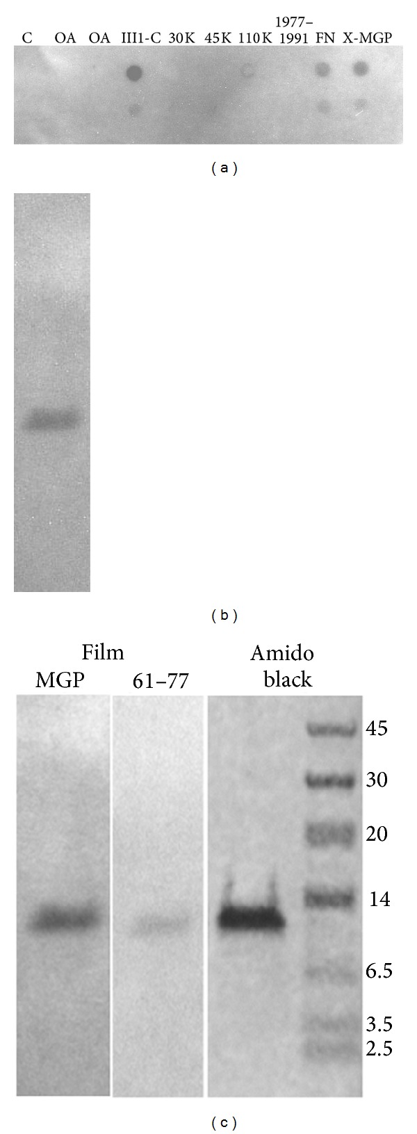
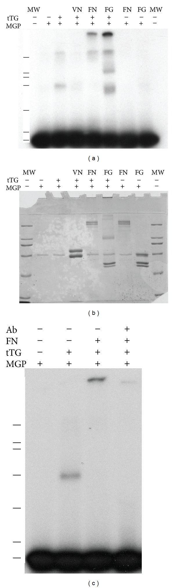
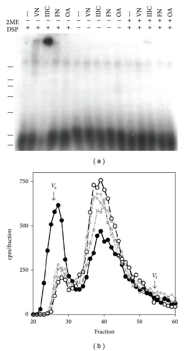
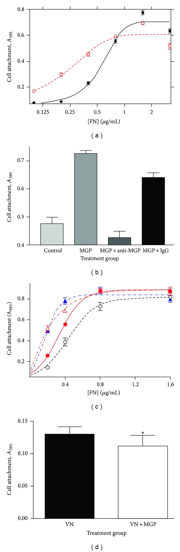
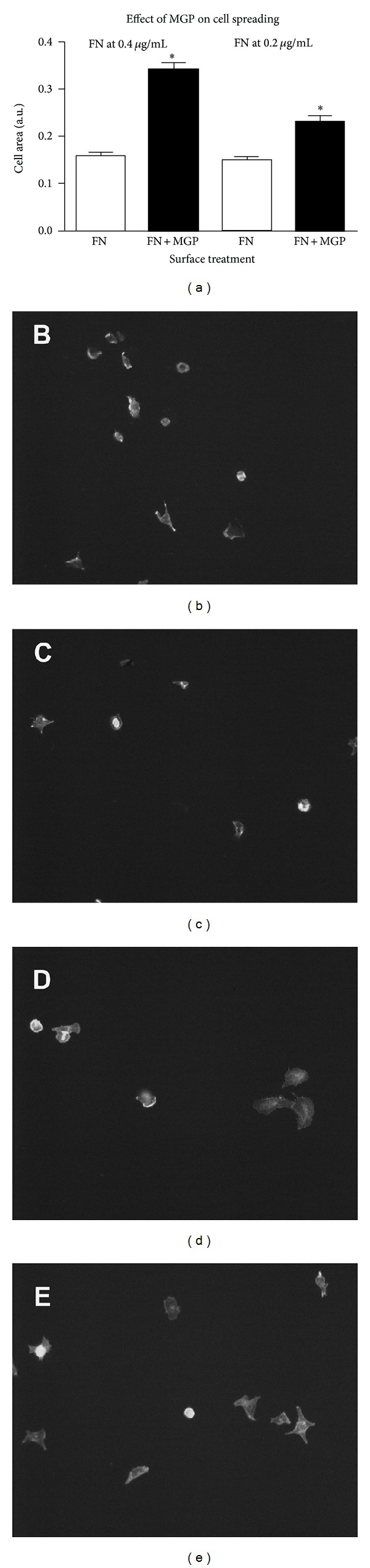
Background. Matrix Gla protein (MGP) is a vitamin K-dependent, extracellular matrix protein. MGP is a calcification inhibitor of arteries and cartilage. However MGP is synthesized in many tissues and is especially enriched in embryonic tissues and in cancer cells. The presence of MGP in those instances does not correlate well with the calcification inhibitory role. This study explores a potential mechanism for MGP to bind to matrix proteins and alter cell matrix interactions. Methods. To determine whether MGP influences cell behavior through interaction with fibronectin, we studied MGP binding to fibronectin, the effect of MGP on fibronectin mediated cell attachment and spreading and immunolocalized MGP and fibronectin. Results. First, MGP binds to fibronectin. The binding site for MGP is in a specific fibronectin fragment, called III1-C or anastellin. The binding site for fibronectin is in a MGP C-terminal peptide comprising amino acids 61-77. Second, MGP enhances cell attachment and cell spreading on fibronectin. MGP alone does not promote cell adhesion. Third, MGP is present in fibronectin-rich regions of tissue sections. Conclusions. MGP binds to fibronectin. The presence of MGP increased cell-fibronectin interactions.


Figures

Figure 1
Figure 1
MGP binds to fibronectin and to the first type III repeat domain of fibronectin III1-C. (a) MGP binds fibronectin and fibronectin fragments with type III repeats. Dot blot spots with radioactive MGP probe and 100 microliters of protein solutions were dot blotted onto the nitrocellulose filter, then blocked with 1% BSA-PBS, overlaid with radioactive MGP, washed, and exposed to film as described in methods. C, PBS buffer; OA, 1 mg/mL ovalbumin; FNIII1C, 1 mg/mL fibronectin III1-C; 30 KFN, 1 mg/mL 30 kDA fragment of fibronectin; 45 KFN, 1 mg/mL 45 kDA fragment of fibronectin; 110 KFN, 1 mg/mL 110 kDA fragment of fibronectin; 1991–1997, 1 mg/mL of the cell binding peptide of fibronectin comprised of amino acids 1991 through 1997; FN, 1 mg/mL fibronectin; anti-MGP, 5 μg/mL of anti-MGP IgG from rabbit. The lower row of dot blotted proteins contains 10x lower concentration of proteins. (b) Radioactive MGP probe binds to fibronectin III1-C on far western blot. Fibronectin III1-C is run on SDS-PAGE, electroblotted, blocked and overlaid with iodinated MGP, washed, and exposed to film as described in methods. (c) Both MGP and the 61–77 C-terminal peptide of MGP bind to fibronectin III1-C. Far western blot with either 125I-MGP or 125I-MGP61–77 overlay on fibronectin III1-C and exposed to film as described. Both probes bind to a band that comigrates with a gel band of fibronectin III1-C shown stained with amido black next to molecular weight markers.
Figure 2
Figure 2
MGP is incorporated into crosslinked fibronectin and fibrinogen by tissue transglutaminase. SDS-PAGE of reaction products of MGP, tTG, and extracellular matrix proteins. Lines indicate 7 marker positions of 200, 116, 97, 66, 43, 31, and 14 kDa. (a) Autoradiograph of 7.5% acrylamide SDS-PAGE gel the legend above indicates whether 125I-labeled MGP or tTG was present as + and − symbols; The protein incubated with MGP is shown in the top line, VN, vitronectin, FN, fibronectin, and FG, fibrinogen. MW indicates that molecular weight markers were run in those lanes. (b) Coomassie blue stained 7.5% acrylamide SDS-PAGE gel used for autoradiograph in (a). Molecular weight markers are 200, 116, 97, 66, 43, 31, and 14 kDa. MGP runs with the 14 kDa protein at the dye front on this gel. (c) Autoradiograph of 7.5% SDS-PAGE gel. The furthest right lane marked Ab+ shows inhibitory effect of added anti-MGP IgG on incorporation of MGP into crosslinked fibronectin.
Figure 3
Figure 3
MGP is crosslinked by DSP to vitronectin, fibronectin, and fibronectin III1-C polypeptide. (a) SDS-PAGE of DSP crosslinking reaction products of unlabeled proteins incubated with trace amounts of radioactive MGP. The presence of the crosslinker DSP, reducing agent 2-mercaptoethanol (2ME), is indicated by + or − in the appropriate lane. The protein present in the reaction is indicated above the lane; −, no added protein; VN, vitronectin; IIIC fibronectin III1-C polypeptide; FN, fibronectin; and OA, ovalbumin. (b) Gel filtration column separation of DSP crosslinked MGP and Fibronectin III1-C. Lines indicate 7 marker positions of 200, 116, 97, 66, 43, 31, and 14 kDa. DSP reacted sample containing labeled MGP and unlabeled III1-C (●); DSP reacted solution containing labeled MGP and unlabeled III1-C reduced with mercaptoethanol prior to column loading, (◯); labeled MGP alone (); Labeled MGP and unlabeled III1-C incubated with DSP vehicle (DMSO) (◊); DSP reacted solution containing labeled MGP alone (□). The exclusion volume V e was estimated by blue dextran elution, and total volume V t was estimated by phenol red elution. The column was run with 4 M guanidine containing buffer to disassociate noncovalent interactions (see methods).
Figure 4
Figure 4
MGP enhances cell attachment to fibronectin. (a) The y-axis is HeLa cell binding indicated as absorbance at 595 nm. The x-axis is the log of fibronectin concentration from 0 to 3.3 μg/mL either with 3 μg/mL MGP (◯) or control buffer (●). MGP did not enhance cell attachment for 0 fibronectin (not shown, logarithmic plot). (b) Anti-MGP IgG blocks the activity of MGP for enhancing cell attachment to fibronectin. Cell attachment was measured for control, 0.4 μg/mL fibronectin; MGP, 0.4 μg/mL fibronectin plus 3 μg/mL MGP; MGP + anti MGP, μg/mL fibronectin plus 3 μg/mL MGP plus 31 μg/mL anti-MGP IgG; and MGP + IgG, 0.4 μg/mL fibronectin plus μg/mL MGP plus 31 μg/mL rabbit polyclonal IgG. MGP and MGP + IgG were significantly different from control and MGP + antiMGP (P ≤ .05). (c) The concentration of MGP enhances cell attachment in a dose responsive manner but does not change maximum cell binding to fibronectin. Experiments were performed as in (a) except that MGP concentrations of 0.0, 0.8, 3, and 6 are compared, and the highest concentration of fibronectin is 1.6 μg/mL. No MGP (◯); 0.8 μg/mL MGP (●); 3 μg/mL MGP (); and 6 μg/mL MGP (▲). Error bars are SEM for 4–6 replicate binding assays for each data point. Lines are nonlinear regression lines derived by Prism software. (d) MGP inhibits cell binding to vitronectin. VN, 0.2 μg/mL vitronectin; VN + MGP, 0.2 μg/mL vitronectin plus 0.8 μg/mL MGP. Error Bars are SEM for 8 replicate binding assays. The ∗ indicates significant difference (P < .05).
Figure 5
Figure 5
MGP augments cell spreading on fibronectin. In Panel (a) the graph shows the calculated cell area/cell for fibronectin coated surfaces compared to fibronectin plus MGP coated surfaces. FN is fibronectin alone open bars at 0.4 μg/mL (left) or 0.2 μg/mL (right); FN + MGP dark bars is the indicated concentration of fibronectin plus MGP at 3 μg/mL. The asterisk indicates that the combination of FN + MGP was significantly different from FN at each concentration of FN (P ≤ .0001, error bars are SEM). The average cell area from a minimum of 100 cells in 14 to 16 random microscopic fields is shown. Cells were allowed to attach for 2 hours in serum-free medium then fixed, stained, and imaged and the cell area quantified. Area per cell was determined by NIH Image software as described in experimental procedures. Panels (b) and (c) are images of cells attached to fibronectin alone (0.4 μg/mL), and Panels (d) and (e) are images of cells attached to fibronectin plus MGP (0.4 μg/mL and 3 μg/mL, resp.).
Figure 6
Figure 6
MGP and fibronectin localization in the embryonic rat kidney. (a)–(c) serial section of E20 rat kidney in which fibronectin is localized with mouse-anti-fibronectin and Vectastain ABC elite universal peroxidase-diaminobenzidine and counterstained with methyl green. (d)–(f) Serial section of E20 rat kidney in which MGP localized with rabbit-anti-MGP, Vectastain ABC elite universal peroxidase diaminobenzidine and counterstained with methyl green. A and D are low power images of the same region of kidney taken with the 6.3x objective. U is the lumen of the developing ureter, and G is a developing glomerulus. Fibronectin localized in B and C taken with a 40x oil immersion objective. U is the lumen of the developing ureter. Arrows indicate tubules identical to those shown in the serial section. MGP localized in E and F, image of equivalent fields as B and C, taken at the same magnification with a 40x objective. U is the developing ureter. Arrows indicate intensely stained tubules for comparison.

Similar articles

Cited by

References

    1. Fraser JD, Price PA. Lung, heart, and kidney express high levels of mRNA for the vitamin K-dependent matrix Gla protein. Implications for the possible functions of matrix Gla protein and for the tissue distribution of the γ-carboxylase. The Journal of Biological Chemistry. 1988;263(23):11033–11036. - PubMed
    1. Price PA, Rice JS, Williamson MK. Conserved phosphorylation of serines in the Ser-X-Glu/Ser(P) sequences of the vitamin K-dependent matrix Gla protein from shark, lamb, rat, cow, and human. Protein Science. 1994;3(5):822–830. - PMC - PubMed
    1. Zhao J, Araki N, Nishimoto SK. Quantitation of matrix Gla protein mRNA by competitive polymerase chain reaction using glyceraldehyde-3-phosphate dehydrogenase as an internal control. Gene. 1995;155(2):159–165. - PubMed
    1. Zhao J, Nishimoto SK. Matrix Gla protein gene expression is elevated during postnatal development. Matrix Biology. 1996;15(2):131–140. - PubMed
    1. Luo G, Ducy P, McKee MD, et al. Spontaneous calcification of arteries and cartilage in mice lacking matrix GLA protien. Nature. 1997;386(6620):78–81. - PubMed

LinkOut - more resources