Review
. 2014 Jun 11;10(16):23-32.
doi: 10.1016/j.csbj.2014.05.003. eCollection 2014 Jun.

Structure and function of nucleotide sugar transporters: Current progress

Affiliations
Review

Structure and function of nucleotide sugar transporters: Current progress

Barbara Hadley et al. Comput Struct Biotechnol J. .

Abstract

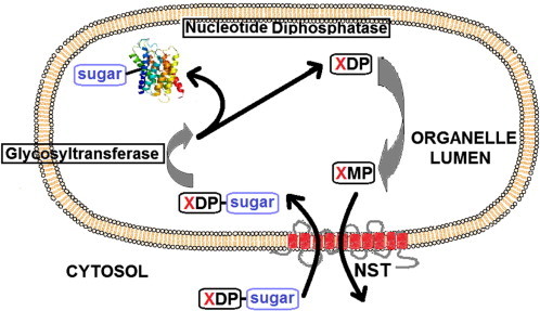
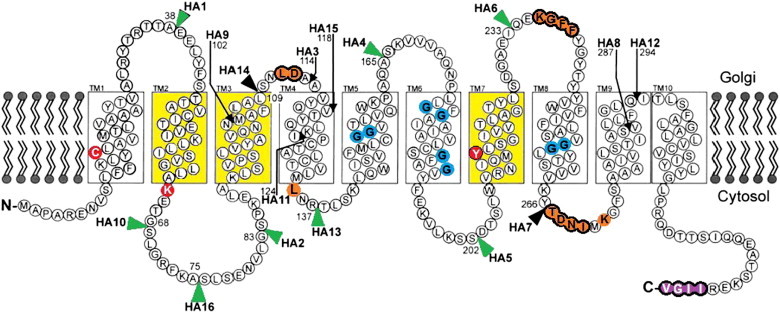
The proteomes of eukaryotes, bacteria and archaea are highly diverse due, in part, to the complex post-translational modification of protein glycosylation. The diversity of glycosylation in eukaryotes is reliant on nucleotide sugar transporters to translocate specific nucleotide sugars that are synthesised in the cytosol and nucleus, into the endoplasmic reticulum and Golgi apparatus where glycosylation reactions occur. Thirty years of research utilising multidisciplinary approaches has contributed to our current understanding of NST function and structure. In this review, the structure and function, with reference to various disease states, of several NSTs including the UDP-galactose, UDP-N-acetylglucosamine, UDP-N-acetylgalactosamine, GDP-fucose, UDP-N-acetylglucosamine/UDP-glucose/GDP-mannose and CMP-sialic acid transporters will be described. Little is known regarding the exact structure of NSTs due to difficulties associated with crystallising membrane proteins. To date, no three-dimensional structure of any NST has been elucidated. What is known is based on computer predictions, mutagenesis experiments, epitope-tagging studies, in-vitro assays and phylogenetic analysis. In this regard the best-characterised NST to date is the CMP-sialic acid transporter (CST). Therefore in this review we will provide the current state-of-play with respect to the structure-function relationship of the (CST). In particular we have summarised work performed by a number groups detailing the affect of various mutations on CST transport activity, efficiency, and substrate specificity.

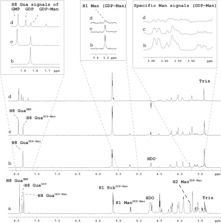
Keywords: CMP-sialic acid transporter; Endoplasmic reticulum; Golgi apparatus; Nucleotide sugar transporters; STD NMR spectroscopy.


Figures

Fig. 1
Fig. 1
The general transport mechanism of NSTs. The XDP-sugar (nucleotide sugar donor) enters the lumen of the organelle in exchange for the corresponding nucleoside monophosphate (XMP). After entering the lumen the sugar is transferred to either a protein or lipid in a reaction catalysed by glycosyltransferases. The diphosphate nucleotide (XDP) is then acted upon by a membrane-bound nucleotide diphosphatase producing the XMP that is subsequently exported . In some cases where the nucleotide sugar donor is a monophosphate, the dephosphorylation reaction performed by the diphosphatase is not required.
Fig. 2
Fig. 2
The direct analysis of Aspergillus GMT interaction with GDP-Man, GDP and GMP using STD NMR spectroscopy. 1H (a) and competition STD NMR spectra of Aspergillus Golgi-enriched fractions complexed with GDP-Man (b) followed by the addition of equimolar amounts of GMP (c) and GDP (d). Some STD signals were found to increase due to overlapping chemical shifts (e.g. the H1 ribose signal at 5.65 ppm), however the H8 guanine signal of the three ligands does not have the same chemical shift and therefore could be used to monitor the interaction of the GMT with GDP-Man, GDP and GMP. The H8 GuaGDP-Man signal (b) is reduced following the addition of GMP and GDP (c and d, respectively) with a corresponding appearance of H8 guanine signals associated with GMP and GDP. Specific mannose signals were reduced by ∼ 50% following the addition of equimolar GMP (c), and the signals after addition of GDP (d) showed a further reduction of ∼ 50% compared to (c).
Fig. 3
Fig. 3
Diagram representing the membrane topology of CST as proposed by independent studies. 1. TM1–TM10 were identified using HA-epitope tagging . The position of HA epitopes used to deduce this model is indicated by arrows and arrowheads. Black arrows and arrowheads indicate HA tags that inactivated CST, whereas the green arrowheads mark the position of HA tags that did not inactivate the CST. 2. The TM domains coloured in yellow are essential for CST activity as identified through UGT–CST chimeras . When TM2, TM3 and TM7 from CST were engineered into UGT, the resulting transporter was then able to transport both CMP-Sia and UDP-Gal. 3. Deletion of the four purple coloured amino acids eliminated the export signals and prevented ER to Golgi translocation 4. The blue coloured Gly residues were identified as contributing to the formation of a putative aqueous channel necessary for the translocation of CMP-Sia . 5. The orange coloured amino acids ringed in black were identified by GFP-tagging as essential for CST activity. The orange amino acids with no black ring were identified as essential by point mutations . 6. Amino acids in red were identified as being essential for CST substrate recognition . Diagram modified from Eckhardt, Gotza & Gerardy-Schahn (1999) and Maggioni, Martinez-Duncker & Tiralongo (2013) .

References

    1. Shental-Bechor D., Levy Y. Effect of glycosylation on protein folding: a close look at thermodynamic stabilization. Proc Natl Acad Sci U S A. 2008;105:8256–8261. - PMC - PubMed
    1. Hayashi H., Yamashita Y. Role of N-glycosylation in cell surface expression and protection against proteolysis of the intestinal anion exchanger SLC26A3. Am J Physiol Cell Physiol. 2012;302:C781–C795. - PubMed
    1. van Kooyk Y., Rabinovich G.A. Protein-glycan interactions in the control of innate and adaptive immune responses. Nat Immunol. 2008;9:593–601. - PubMed
    1. Zhao Y.Y., Takahashi M., Gu J.G., Miyoshi E., Matsumoto A., Kitazume S. Functional roles of N-glycans in cell signaling and cell adhesion in cancer. Cancer Sci. 2008;99:1304–1310. - PMC - PubMed
    1. Zhou F., Xu W., Hong M., Pan Z., Sinko P.J., Ma J. The role of N-linked glycosylation in protein folding, membrane targeting, and substrate binding of human organic anion transporter hOAT4. Mol Pharmacol. 2005;67:868–876. - PubMed