

Spontaneous resolution of a fetal dural sinus thrombosis: one case report and review of the literatures
- PMID: 25210612
- PMCID: PMC4152190
Spontaneous resolution of a fetal dural sinus thrombosis: one case report and review of the literatures
Abstract
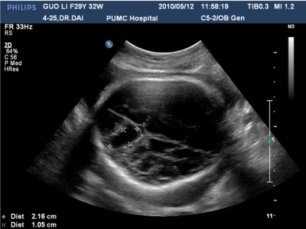
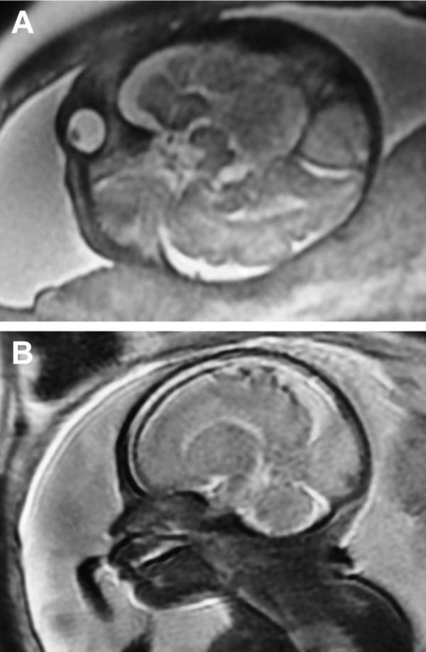
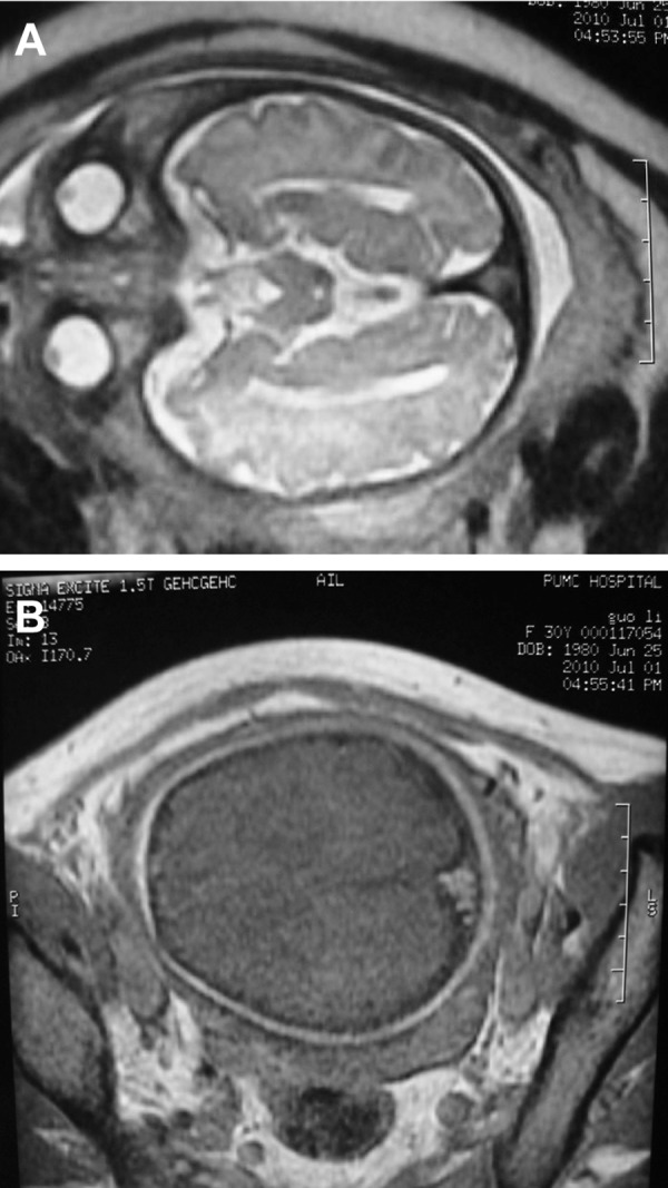
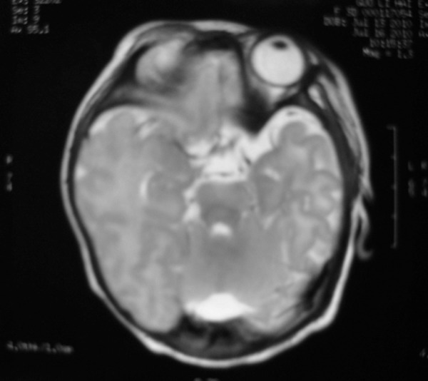
Fetal dural sinus thrombosis is a rare finding. Most cases have been terminated without long-term follow-ups. Recently some reports have indicated the potentially favorable evolution of fetal dural sinus thrombosis. Most of the fetuses showing symptoms have been delivered with normal neurologic outcome. We report a case of fetal dural sinus thrombosis. Serial ultrasound and magnetic resonance images (MRI) showed the shrinkage of the thrombosis which indicated good prognosis. No physical or neurological abnormality was observed at 8-months follow-up. Conservative treatment is appropriate to prenatally diagnosed dural sinus thrombosis with favorable prognostic factors. Serial MRI or ultrasound should be taken every 1-2 months to monitor the thrombosis development and fetal well-beings.
Keywords: Fetus; Intracranial; Prenatal Diagnosis; Sinus Thrombosis.
Figures
Similar articles
-
Prenatal diagnosis of thrombosis of the dural sinuses: report of six cases, review of the literature and suggested management.Ultrasound Obstet Gynecol. 2008 Aug;32(2):188-98. doi: 10.1002/uog.5348. Ultrasound Obstet Gynecol. 2008. PMID: 18512853 Review.
-
In utero magnetic resonance imaging for diagnosis of dural venous sinus ectasia with thrombosis in the fetus.Pediatr Radiol. 2013 Dec;43(12):1591-8. doi: 10.1007/s00247-013-2745-7. Epub 2013 Oct 15. Pediatr Radiol. 2013. PMID: 24127016
-
Spontaneous resolution of prenatally diagnosed dural sinus thrombosis: a case report.Ultrasound Obstet Gynecol. 2006 May;27(5):562-5. doi: 10.1002/uog.2764. Ultrasound Obstet Gynecol. 2006. PMID: 16586467 Review.
-
Fetal dural sinus malformation: A case report and discussion of the literature.Australas J Ultrasound Med. 2021 Oct 5;24(4):249-252. doi: 10.1002/ajum.12285. eCollection 2021 Nov. Australas J Ultrasound Med. 2021. PMID: 34888135 Free PMC article.
-
Thrombosed Fetal Dural Sinus Malformation: A Case Report.J Pharm Bioallied Sci. 2024 Dec;16(Suppl 4):S4145-S4148. doi: 10.4103/jpbs.jpbs_894_24. Epub 2024 Oct 30. J Pharm Bioallied Sci. 2024. PMID: 39927033 Free PMC article.
Cited by
-
Letter to the Editor Regarding 'A neonatal case of cerebral venous sinus thrombosis with intrauterine onset after COVID19 infection during pregnancy: cause or coincidence?'.J Stroke Cerebrovasc Dis. 2023 May;32(5):107033. doi: 10.1016/j.jstrokecerebrovasdis.2023.107033. J Stroke Cerebrovasc Dis. 2023. PMID: 37030970 Free PMC article. No abstract available.
References
-
- Gicquel JM, Potier A, Sitruk S, Girard N. Normal outcome after prenatal diagnosis of thrombosis of the torcular herophili. Prenat Diagn. 2000;20(10):824–827. - PubMed
-
- Visentin A, Falco P, Pilu G, Perolo A, Valeri B, Santini D, et al. Prenatal diagnosis of thrombosis of the dural sinuses with real-time and color Doppler ultrasound. Ultrasound Obstet Gynecol. 2001;17(4):322–325. - PubMed
-
- Emamian SA, Bulas DI, Vezina GL, Dubovskyi EC, Cogan P. Fetal MRI evalution of an intracranial mass: in utero evalution of hemorrhage. Pediatr Radiol. 2002;32(8):593–597. - PubMed
-
- Clode N, Cardoso C, Tavares J, Albuquerque M, Silva JL, Graca LM. Prenatal diagnosis of thrombosis of dural sinus. Ultrasound Obstet Gynecol. 2004;24(3):330–330.
-
- Jung E, Won HS, Kim SK, Shim JY, Lee PR, Kim A, et al. Spontaneous resolution of prenatally diagnosed dural sinus thrombosis: a case report. Ultrasound Obstet Gynecol. 2006;27(5):562–565. - PubMed
LinkOut - more resources
Full Text Sources