. 2014 Aug 28:5:28.
doi: 10.1186/2041-9139-5-28. eCollection 2014.

The role of a lens survival pathway including sox2 and αA-crystallin in the evolution of cavefish eye degeneration

Affiliations

The role of a lens survival pathway including sox2 and αA-crystallin in the evolution of cavefish eye degeneration

Li Ma et al. Evodevo. .

Abstract

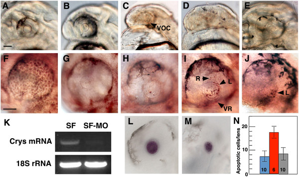
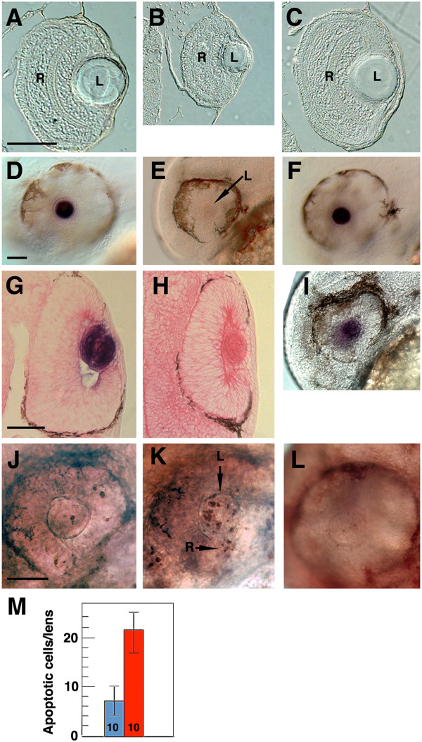
Background: The teleost Astyanax mexicanus is a single species consisting of eyed surface-dwelling (surface fish) and blind cave-dwelling (cavefish) morphs. Cavefish eyes are lost through apoptosis of the lens, which in turn promotes the degeneration of other optic tissues. The αA-crystallin (αA-crys) gene is strongly downregulated in the cavefish lens and is located in a genomic region (QTL) responsible for eye loss. Therefore, αA-crys has been proposed as a candidate for regulating cavefish eye degeneration. The purpose of this study was to determine the mechanism of αA-crys downregulation and its role in cavefish eye degeneration.

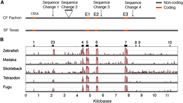
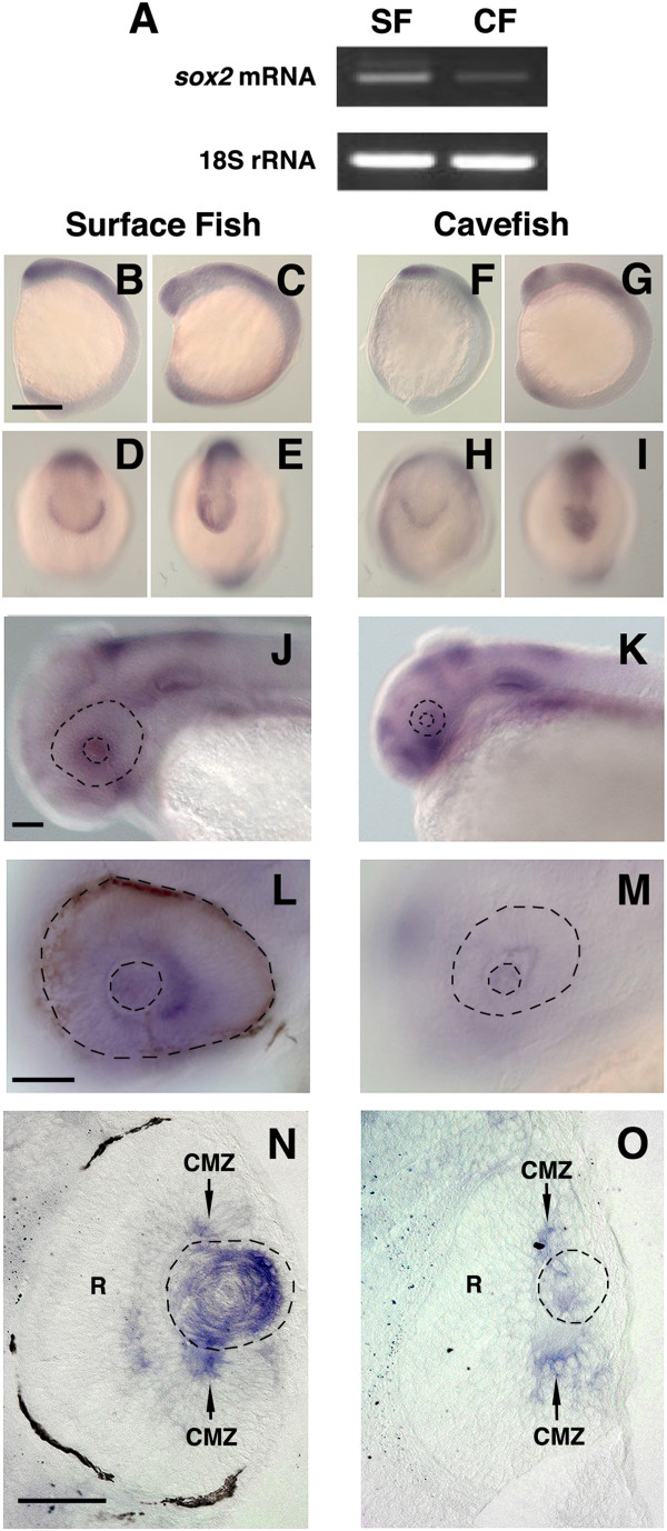
Results: The involvement of αA-crys in eye degeneration was confirmed by knocking down its expression in surface fish, which led to apoptosis of the lens. The underlying reason for αA-crys downregulation in cavefish was investigated by comparing genomic αA-crys DNA sequences in surface fish and cavefish, however, no obvious cis-regulatory factors were discovered. Furthermore, the cavefish αA-crys allele is expressed in surface fish x cavefish F1 hybrids, indicating that evolutionary changes in upstream genes are most likely responsible for αA-crys downregulation. In other species, Sox2 is one of the transcription factors that regulate lens crystallin genes during eye development. Determination of sox2 expression patterns during surface fish and cavefish development showed that sox2 is specifically downregulated in the cavefish lens. The upstream regulatory function of Sox2 was demonstrated by knockdown in surface fish, which abolished αA-crys expression and induced lens apoptosis.

Conclusions: The results suggest that αA-crys is required for normal eye development in cavefish via suppression of lens apoptosis. The regulatory changes involved in αA-crys downregulation in cavefish are in trans-acting factors rather than cis-acting mutations in the αA-crys gene. Therefore, αA-crys is unlikely to be the mutated gene(s) associated with an Astyanax eye QTL. The results reveal a genetic pathway leading from sox2 to αA-crys that is required for survival of the lens in Astyanax surface fish. Defects in this pathway may be involved in lens apoptosis and thus a cause of cavefish eye degeneration.

Keywords: Astyanax mexicanus; Blind cavefish; Cis and trans gene regulation; Eye degeneration; Lens apoptosis; Lens survival pathway; Sox2; αA-crystallin.


Figures

Figure 1
Figure 1
αA-crys knockdown with morpholinos (MOs). (A-E) Effects of MO on lens development. (A) Embryos injected with control MO have a normal sized lens. (B-D) Embryos injected with αA-crys splice-blocking morpholinos (sbMO) and translation-blocking morpholino (tbMO) develop a normal sized lens (B), a lens of reduced size and a reduced ventral optic cup (VOC) (C)1, or no lens (D). (E) Embryos injected with αA-crys sbMO and αA-crys mRNA show normal lens development. (F-J) Apoptosis detected by terminal deoxynucleotidyl transferase dUTP nick end labeling (TUNEL). (F) An uninjected embryo treated with DNase shows apoptotic cells throughout the eye. (G, H) Uninjected (G) and control MO-injected (H) embryos show background levels of lens apoptosis. (I) Embryos injected with αA-crys sbMO and tbMO embryos show apoptotic cells in the lens (L) and retina (R). VR: reduced ventral retina. (J) Embryos injected with αA-crys sbMO and αA-crys mRNA show background levels of lens apoptosis. (K) Semiquantitative RT-PCR . SF: uninjected embryos. SF-MO: αA-crys MO-injected embryos. (L, M)In situ hybridization shows mip gene expression in the lens of uninjected (L) and αA-crys sbMO-injected (M) embryos. All embryos are shown at 40 hr post fertilization (hpf). All scale bars are 80 μm; A-E, F-J, L and M are the same magnifications. (N) Histogram showing lens apoptotic cells in control MO-injected embryos (blue), αA-crys sbMO-injected embryos (red), and αA-crys sbMO and αA-crys mRNA-injected embryos (gray). Error bars represent SD. Numbers at the base of the histogram represent sample sizes. P = 0.00 (one-way analysis of variance (ANOVA).
Figure 2
Figure 2
Sequence changes in the surface fish and cavefish αA-crys gene loci. A. A schematic diagram of the sequenced Texas surface fish (SF) and Pachón cavefish (CF) αA-crys loci showing the positions of four sequence changes (see Table 1). CßSA: last cystathionine-ß-synthase a exon. E1-3: αA-crys exons. B. Alignment of αA-crys loci in 5 teleost species with Astyanax Texas surface fish αA-crys shows 10 conserved regions (black bars labeled 1 to 10 on top of frame) but their sequences are not changed between surface fish and cavefish (Table 2).
Figure 3
Figure 3
PCR amplification of genomic DNA yields a 1,003-bp amplicon containing the 633-bp region (sequence change 2 in Figure2A) in Mexican (RC) surface fish (SF) and Pachón (Pa), Los Sabinos (LS), Tinaja (Ti), Jineo (Ji), Chica (Ch), and Molino (Mo) cavefish populations and a 370-bp amplicon lacking the 633-bp region in the Texas (Tx) surface fish population. PCR amplification was carried out using primers flanking the 633-bp region in Pachón cavefish (see Methods).
Figure 4
Figure 4
αA-crys expression is controlled by trans-acting factors in F1 hybrid embryos. (A-D)In situ hybridization showing αA-crys expression in the lens of (A) surface fish (SF), (B) surface fish X cavefish (CF) F1 hybrids, and (C) cavefish X surface fish F1 hybrids, but not in (D) cavefish embryos at 72 hr post fertilization (hpf). The eye and lens are outlined by dashes in cavefish (D). Scale bar in A is 150 μm; magnifications are the same in A-D. (E) Semiquantitative RT-PCR showing αA-crys transcript levels in 40 hpf surface fish (SF), surface fish x cavefish F1 hybrid embryos (HY), and cavefish (CF) embryos compared to an 18S rRNA standard. (F) Profile of sequenced RT-PCR products from surface fish x cavefish F1 hybrid embryos showing a mixture of A (green) and G (black) residues at the first exon site distinguishing the surface fish from cavefish αA-crys alleles.
Figure 5
Figure 5
sox2 expression is downregulated in the lens during cavefish development. (A) Semiquantitative RT-PCR showing reduced sox2 mRNA levels in cavefish relative to surface fish embryos. (B-O)In situ hybridization shows sox2 expression during surface fish (B-E, J, L, N) and cavefish (F-I, K, M, O) development. B-I Tailbud (B, D, F, H) and 5-somite (C, E, G, I) stages viewed from the lateral (B, C, F, G) or rostral (D, E, H, I) sides. Scale bar in B is 250 μm; magnification is the same in B-I. (J-O)sox2 downregulation in the cavefish lens at 42 hr post fertilization (hpf). N and O are sections through the eye region of the embryos shown in J-M. Dashed lines indicate the eye and lens. R: retina. CMZ: ciliary marginal zone. Scale bar in J, L and N is 100 μm; magnification is the same in J, K; L, M; and N, O.
Figure 6
Figure 6
sox2 knockdown. (A-C) Effects of sox2 MO on eye development at 42 hr post fertilization (hpf). Sections of uninjected (A), sox2 MO injected (B), and control MO-injected (C) embryos show reduced eye size after sox2 knockdown. Scale bar in A is 150 μm: A-C are the same magnifications. (D-H)sox2 knockdown abolishes lens αA-crys gene expression. In situ hybridization of uninjected (D), sox2 MO-injected (E), and control MO-injected (F) embryos shows no lens αA-crys expression after sox2 knockdown. Sections through the eyes of in situ hybridized uninjected (G) and sox2 injected (G) embryos confirm the absence of αA-crys expression in the sox2 morphant lens. R: retina. L: lens. (I)In situ hybridization showing lens mip gene expression in sox2 MO-injected embryos. (J-L) Effects of sox2 gene knockdown on apoptosis at 42 hpf. Terminal deoxynucleotidyl transferase dUTP nick end labeling (TUNEL) assays of un-injected (J), sox2 MO-injected (K), and control MO injected (L) embryos show apoptosis in the lens (arrow labeled L) and retina (arrow labeled R) in sox2 morphants. Embryos are shown at 42 (A-I) and 48 (J-L) hpf. Scale bars in D, G, and J are 100 μm; magnification is the same in A-C; D-F, I; G and H; and J-L. M. Histogram showing apoptotic cells in the lens of uninjected embryos (blue) and sox2 MO-injected embryos (red). Error bars represent SD. Numbers at the base of the histogram represent sample sizes. P = 0.00, one-way analysis of variance (ANOVA).

References

    1. Protas M, Jeffery WR. Evolution and development in cave animals: from fish to crustaceans. WIRES Dev Biol. 2012;1:823–845. doi: 10.1002/wdev.61. - DOI - PMC - PubMed
    1. Jeffery WR. Cavefish as a model system in evolutionary developmental biology. Dev Biol. 2001;231:1–12. doi: 10.1006/dbio.2000.0121. - DOI - PubMed
    1. Borowsky R. Restoring sight in blind cavefish. Curr Biol. 2008;18:R23–R24. doi: 10.1016/j.cub.2007.11.023. - DOI - PubMed
    1. Retaux S, Casane D. Evolution of eye development in the darkness of caves: adaptation, drift, or both? EvoDevo. 2013;4:26. doi: 10.1186/2041-9139-4-26. - DOI - PMC - PubMed
    1. Wilkens H. Evolusion and genetics of epigean and cave Astyanax fasciatus (Characidae, Pisces) Evol Biol. 1988;23:271–367.

LinkOut - more resources