Trabecular-iris circumference volume in open angle eyes using swept-source fourier domain anterior segment optical coherence tomography
- PMID: 25210623
- PMCID: PMC4152933
- DOI: 10.1155/2014/590978
Trabecular-iris circumference volume in open angle eyes using swept-source fourier domain anterior segment optical coherence tomography
Abstract
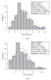
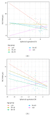
Purpose. To introduce a new anterior segment optical coherence tomography parameter, trabecular-iris circumference volume (TICV), which measures the integrated volume of the peripheral angle, and establish a reference range in normal, open angle eyes. Methods. One eye of each participant with open angles and a normal anterior segment was imaged using 3D mode by the CASIA SS-1000 (Tomey, Nagoya, Japan). Trabecular-iris space area (TISA) and TICV at 500 and 750 µm were calculated. Analysis of covariance was performed to examine the effect of age and its interaction with spherical equivalent. Results. The study included 100 participants with a mean age of 50 (±15) years (range 20-79). TICV showed a normal distribution with a mean (±SD) value of 4.75 µL (±2.30) for TICV500 and a mean (±SD) value of 8.90 µL (±3.88) for TICV750. Overall, TICV showed an age-related reduction (P = 0.035). In addition, angle volume increased with increased myopia for all age groups, except for those older than 65 years. Conclusions. This study introduces a new parameter to measure peripheral angle volume, TICV, with age-adjusted normal ranges for open angle eyes. Further investigation is warranted to determine the clinical utility of this new parameter.
Figures


References
-
- Console JW, Sakata LM, Aung T, Friedman DS, He M. Quantitative analysis of anterior segment optical coherence tomography images: the zhongshan angle assessment program. British Journal of Ophthalmology. 2008;92(12):1612–1616. - PubMed
-
- Liu S, Yu M, Ye C, Lam DSC, Leung CK. Anterior chamber angle imaging with swept-source optical coherence tomography: an investigation on variability of angle measurement. Investigative Ophthalmology and Visual Science. 2011;52(12):8598–8603. - PubMed
-
- Radhakrishnan S, See J, Smith SD, et al. Reproducibility of anterior chamber angle measurements obtained with anterior segment optical coherence tomography. Investigative Ophthalmology and Visual Science. 2007;48(8):3683–3688. - PubMed
-
- Kim DY, Sung KR, Kang SY, et al. Characteristics and reproducibility of anterior chamber angle assessment by anterior-segment optical coherence tomography. Acta Ophthalmologica. 2011;89(5):435–441. - PubMed
Grants and funding
LinkOut - more resources
Full Text Sources
Other Literature Sources

