. 2014 Apr 3:4:e28680.
doi: 10.4161/cl.28680. eCollection 2014.

Distinct patterns of phosphatidylserine localization within the Rab11a-containing recycling system

Affiliations

Distinct patterns of phosphatidylserine localization within the Rab11a-containing recycling system

Nicholas W Baetz et al. Cell Logist. .

Abstract

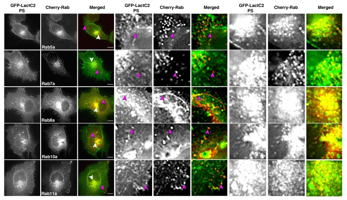
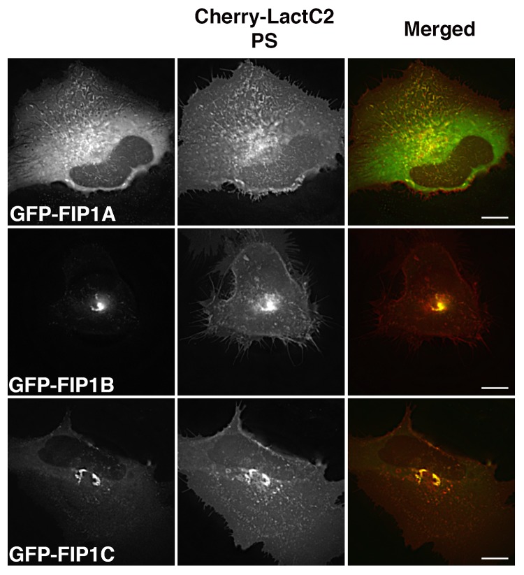
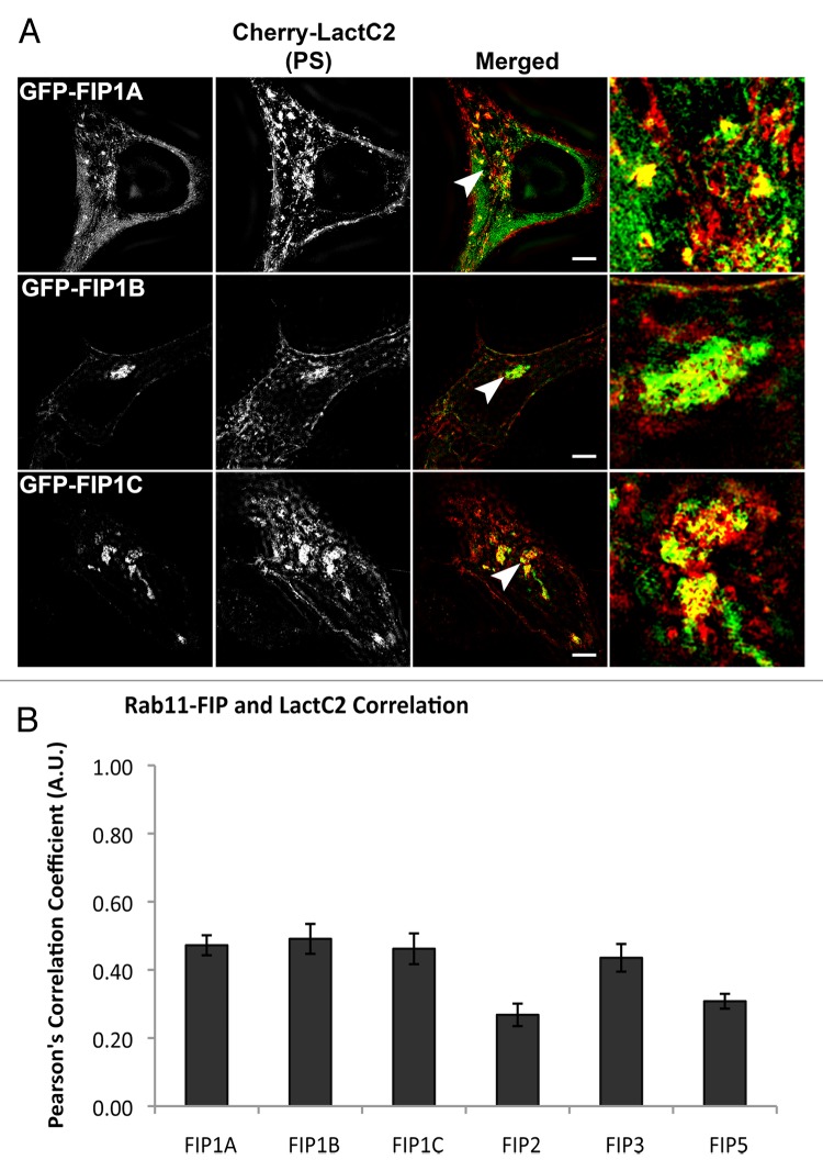
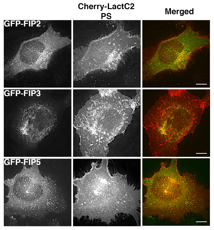
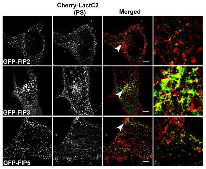
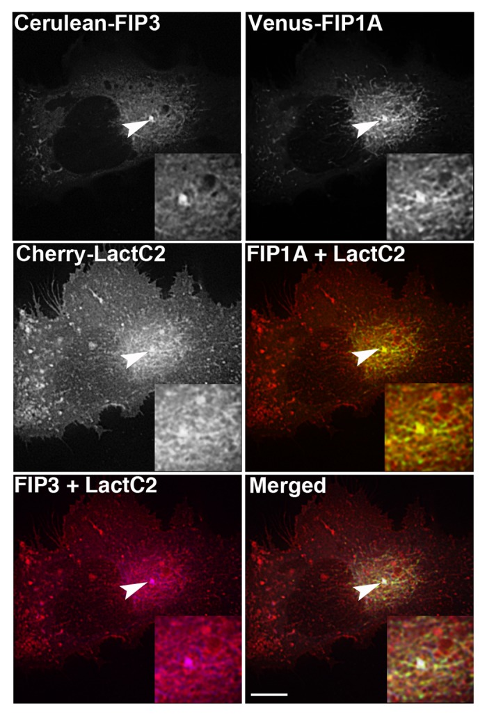
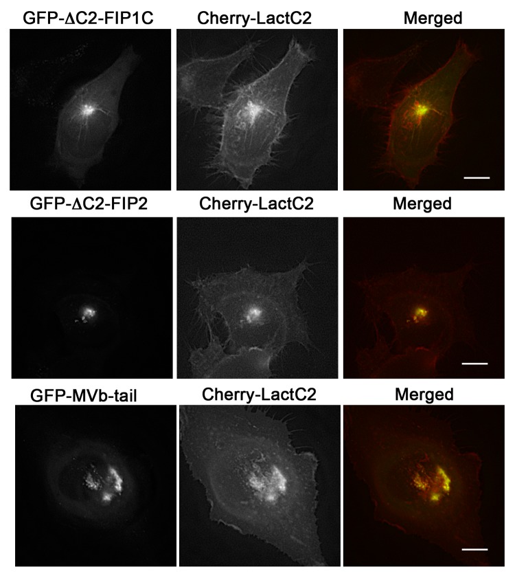
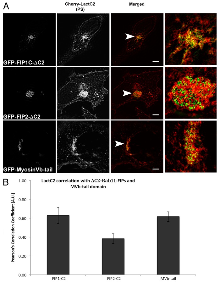
The Rab11 GTPases and Rab11 family-interacting proteins (Rab11-FIPs) define integrated yet distinct compartments within the slow recycling pathway. The lipid content of these compartments is less well understood, although past studies have indicated phosphatidylserine (PS) is an integral component of recycling membranes. We sought to identify key differences in the presence of PS within Rab and Rab11-FIP containing membranes. We used live cell fluorescence microscopy and structured illumination microscopy to determine whether the previously published LactC2 probe for PS displays differential patterns of overlap with various Rab GTPases and Rab11-FIPs. Selective overlap was observed between the LactC2 probe and Rab GTPases when co-expressed in HeLa cells. Rab11-FIP1 proteins consistently overlapped with LactC2 along peripheral and pericentriolar compartments. The specificity of Rab11-FIP1 association with LactC2 was further confirmed by demonstrating that additional Rab11-FIPs (FIP2, FIP3, and FIP5) exhibited selective association with LactC2 containing compartments. Live cell dual expression studies of Rab11-FIPs with LactC2 indicated that PS is enriched along tubular compartments of the Rab11a-dependent recycling system. Additionally, we found that the removal of C2 domains from the Rab11-FIPs induced an accumulation of LactC2 probe in the pericentriolar region, suggesting that inhibition of trafficking through the recycling system can influence the distribution of PS within cells. Finally, we confirmed these findings using structured illumination microscopy suggesting that the overlapping fluorescent signals were on the same membranes. These results suggest distinct associations of Rab GTPases and Rab11-FIPs with PS-containing recycling system membrane domains.

Keywords: Rab11-FIP; Rab11a; Rab5; Rab7; Rab8a; live cell microscopy; phophatidylserine; structured illumination.


Figures

None
Figure 1. EGFP-LactC2 (PS) and mCherry-Rab GTPases in live HeLa cells show distinct patterns of overlap. HeLa cells expressing EGFP-LactC2 and mCherry-Rab proteins were imaged with live cell deconvolution microscopy for at least 1 min every 2 s. Rab5a and Rab7a showed limited overlap with LactC2 in the periphery of cells while Rab8a and Rab11a display consistent overlap with mCherry-LactC2 throughout the cell. Rab10 overlap with LactC2 was present but not consistent over multiple experiments. Data represent at least 3 independent experiments. Bars, 10 μm
None
Figure 2. EGFP-Rab11-FIP1 proteins consistently overlap with mCherry-LactC2 (PS) in live HeLa cells. EGFP-Rab11-FIP1 proteins and mCherry-LactC2 overlapped in peripheral and pericentriolar compartments during imaging of live HeLa cells. FIP1B and FIP1C induced a partial accumulation of LactC2 in the pericentriolar compartments. Cells were imaged for at least one minute every 2 s. Data represent at least 3 independent experiments. Bars, 10 μm.
None
Figure 3. The Rab11-FIP1 proteins are within 100–200nm of LactC2 by SIM. (A) HeLa cells transfected with EGFP-Rab11-FIP1 proteins and mCherry-LactC2 were imaged on coverslips using structured illumination microscopy. Each Rab11-FIP1 protein displayed overlap with LactC2. Images were collected over a 1 μm stack of individual HeLa cells. Bars, 10 μm. (B) Pearson’s correlation coefficients were analyzed for each condition. Rab11-FIP1A (0.472 ± 0.029, n = 14 cells), Rab11-FIP1B (0.491 ± 0.044, n = 9 cells), and Rab11-FIP1C (0.462 ± 0.045, n = 8 cells) had statistically similar overlap with LactC2 (P > 0.05) Results were analyzed using an unpaired, two-tailed, Student’s t test and presented as Mean ± SEM.
None
Figure 4. Alternative EGFP-Rab11-FIPs exhibit selective overlap with mCherry-LactC2. HeLa cells coexpressing EGFP-Rab11-FIPs and mCherry-LactC2 were imaged using live cell deconvolution microscopy. Images were collected every 2 s for at least 1 min. EGFP-Rab11-FIP2 and EGFP-Rab11-FIP5 were separate from mCherry LactC2 particularly in the periphery of HeLa cells. EGFP-Rab11-FIP3 was overlapped with mCherry-LactC2 in the pericentriolar region of the cell on distinct tubular membranes. Data represent at least 3 independent experiments. Bars,10 μm.
Figure 5:
Figure 5:
mCherry-LactC2 with alternative EGFP-Rab11-FIPs is limited to EGFP-FIP3 within the pericentriolar region by SIM. A. EGFP-Rab11-FIPs encoded by genes other than Rab11-FIP1 do not overlap as extensively with LactC2 in HeLa cells. HeLa cells expressing EGFP-Rab11-FIPs and mCherry-LactC2 were imaged on coverslips by structured illumination microscopy. EGFP-Rab11-FIP3 was observed along mCherry-LactC2 positive compartments within the perincentriolar region while EGFP-Rab11-FIP2 and EGFP-Rab11-FIP5 did not overlap with mCherry-LactC2. Bars,10 μm. B. Pearson’s correlation coefficients were analyzed for each condition. Rab11-FIP2 (0.268 ± 0.033, n = 8 cells) and Rab11-FIP5 (0.308 ± 0.022, n = 10 cells) had significantly lower correlation coefficients (P < 0.05) than other Rab11-FIPs. Rab11-FIP3 (0.435 ± 0.041, n = 17 cells; P > 0.05) was not significantly different than Rab11-FIP1 proteins. Results were analyzed using an unpaired, two-tailed, Student’s t test and presented as Mean ± SEM.
None
Figure 6. mCherry-LactC2 is enriched along tubular compartments of the Rab11-FIP network. Cerulean-Rab11-FIP3, Venus-Rab11-FIP1A, and mCherry-Rab11-LactC2 expressed in HeLa cells overlap along endosomal tubules in the pericentriolar region. Images were collected every 3 s for at least 1 min. Bars, 10μm.
None
Figure 7. EGFP-Rab11-FIPs lacking the N-terminal C2 domain induce accumulation of LactC2 in the pericentriolar region. EGFP-Rab11-FIP1CΔC2 or EGFP-Rab11-FIP2ΔC2 cause an accumulation of mCherry-LactC2 in the pericentriolar region of live HeLa cells. Expression of MyosinVb-tail induced a similar accumulation of mCherry-LactC2. Images were collected every 2 s for at least 1 min. Data are representative of 2 independent experiments. Bar, 10μm.
Figure 8:
Figure 8:
The mCherry-LactC2 and Rab11-FIP-ΔC2 proteins exhibit selective overlap in the pericentriolar region. (A) EGFP-Rab11-FIPΔC2 constructs coexpressed with mCherry-LactC2 on coverslips and imaged using structured illumination microscopy demonstrated distinct associations between LactC2 and Rab11-FIP1C or Myosin Vb, but visible separation between LactC2 and Rab11-FIP2 . Bars, 10 μm. (B) ΔC2-Rab11-FIP2 (0.383 ± 0.052, n = 9 cells) and LactC2 (P < 0.05) had significantly lower correlation than ΔC2-Rab11-FIP1C (0.629 ± 0.086, n = 3 cells) or Myosin Vb tail (0.616 ± 0.051, n = 9 cells; P > 0.05). Results were analyzed using an unpaired, two-tailed, Student’s t test and presented as Mean ± SEM.

References

    1. Chavrier P, Parton RG, Hauri HP, Simons K, Zerial M. Localization of low molecular weight GTP binding proteins to exocytic and endocytic compartments. Cell. 1990;62:317–29. doi: 10.1016/0092-8674(90)90369-P. - DOI - PubMed
    1. Mizuno-Yamasaki E, Rivera-Molina F, Novick P. GTPase networks in membrane traffic. Annu Rev Biochem. 2012;81:637–59. doi: 10.1146/annurev-biochem-052810-093700. - DOI - PMC - PubMed
    1. Sönnichsen B, De Renzis S, Nielsen E, Rietdorf J, Zerial M. Distinct membrane domains on endosomes in the recycling pathway visualized by multicolor imaging of Rab4, Rab5, and Rab11. J Cell Biol. 2000;149:901–14. doi: 10.1083/jcb.149.4.901. - DOI - PMC - PubMed
    1. Rink J, Ghigo E, Kalaidzidis Y, Zerial M. Rab conversion as a mechanism of progression from early to late endosomes. Cell. 2005;122:735–49. doi: 10.1016/j.cell.2005.06.043. - DOI - PubMed
    1. Di Paolo G, De Camilli P. Phosphoinositides in cell regulation and membrane dynamics. Nature. 2006;443:651–7. doi: 10.1038/nature05185. - DOI - PubMed

LinkOut - more resources