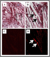
Unique antibody responses to malondialdehyde-acetaldehyde (MAA)-protein adducts predict coronary artery disease
- PMID: 25210746
- PMCID: PMC4161424
- DOI: 10.1371/journal.pone.0107440
Unique antibody responses to malondialdehyde-acetaldehyde (MAA)-protein adducts predict coronary artery disease
Abstract
Malondialdehyde-acetaldehyde adducts (MAA) have been implicated in atherosclerosis. The purpose of this study was to investigate the role of MAA in atherosclerotic disease. Serum samples from controls (n = 82) and patients with; non-obstructive coronary artery disease (CAD), (n = 40), acute myocardial infarction (AMI) (n = 42), or coronary artery bypass graft (CABG) surgery due to obstructive multi-vessel CAD (n = 72), were collected and tested for antibody isotypes to MAA-modifed human serum albumin (MAA-HSA). CAD patients had elevated relative levels of IgG and IgA anti-MAA, compared to control patients (p<0.001). AMI patients had a significantly increased relative levels of circulating IgG anti-MAA-HSA antibodies as compared to stable angina (p<0.03) or CABG patients (p<0.003). CABG patients had significantly increased relative levels of circulating IgA anti-MAA-HSA antibodies as compared to non-obstructive CAD (p<0.001) and AMI patients (p<0.001). Additionally, MAA-modified proteins were detected in the tissue of human AMI lesions. In conclusion, the IgM, IgG and IgA anti-MAA-HSA antibody isotypes are differentially and significantly associated with non-obstructive CAD, AMI, or obstructive multi-vessel CAD and may serve as biomarkers of atherosclerotic disease.
Conflict of interest statement
Figures


Similar articles
-
Malondialdehyde-Acetaldehyde Adducts and Antibody Responses in Rheumatoid Arthritis-Associated Interstitial Lung Disease.Arthritis Rheumatol. 2019 Sep;71(9):1483-1493. doi: 10.1002/art.40900. Epub 2019 Jul 22. Arthritis Rheumatol. 2019. PMID: 30933423 Free PMC article.
-
Autoantibodies to Malondialdehyde-Acetaldehyde Are Detected Prior to Rheumatoid Arthritis Diagnosis and After Other Disease Specific Autoantibodies.Arthritis Rheumatol. 2020 Dec;72(12):2025-2029. doi: 10.1002/art.41424. Epub 2020 Oct 15. Arthritis Rheumatol. 2020. PMID: 32621635 Free PMC article.
-
Malondialdehyde-acetaldehyde adducts and anti-malondialdehyde-acetaldehyde antibodies in rheumatoid arthritis.Arthritis Rheumatol. 2015 Mar;67(3):645-55. doi: 10.1002/art.38969. Arthritis Rheumatol. 2015. PMID: 25417811 Free PMC article.
-
Aldehyde-modified proteins as mediators of early inflammation in atherosclerotic disease.Free Radic Biol Med. 2015 Dec;89:409-18. doi: 10.1016/j.freeradbiomed.2015.09.003. Epub 2015 Nov 4. Free Radic Biol Med. 2015. PMID: 26432980 Review.
-
Role of malondialdehyde-acetaldehyde adducts in liver injury.Free Radic Biol Med. 2002 Feb 15;32(4):303-8. doi: 10.1016/s0891-5849(01)00742-0. Free Radic Biol Med. 2002. PMID: 11841919 Review.
Cited by
-
Novel predictor markers for early differentiation between transient tachypnea of newborn and respiratory distress syndrome in neonates.Int J Immunopathol Pharmacol. 2021 Jan-Dec;35:20587384211000554. doi: 10.1177/20587384211000554. Int J Immunopathol Pharmacol. 2021. PMID: 33722097 Free PMC article.
-
Autoantibodies and B Cells: The ABC of rheumatoid arthritis pathophysiology.Immunol Rev. 2020 Mar;294(1):148-163. doi: 10.1111/imr.12829. Epub 2019 Dec 16. Immunol Rev. 2020. PMID: 31845355 Free PMC article. Review.
-
Plasma Metabolome Normalization in Rheumatoid Arthritis Following Initiation of Methotrexate and the Identification of Metabolic Biomarkers of Efficacy.Metabolites. 2021 Nov 30;11(12):824. doi: 10.3390/metabo11120824. Metabolites. 2021. PMID: 34940582 Free PMC article.
-
Malondialdehyde-Acetaldehyde Modified (MAA) Proteins Differentially Effect the Inflammatory Response in Macrophage, Endothelial Cells and Animal Models of Cardiovascular Disease.Int J Mol Sci. 2021 Nov 30;22(23):12948. doi: 10.3390/ijms222312948. Int J Mol Sci. 2021. PMID: 34884754 Free PMC article.
-
The Translation and Commercialisation of Biomarkers for Cardiovascular Disease-A Review.Front Cardiovasc Med. 2022 Jun 2;9:897106. doi: 10.3389/fcvm.2022.897106. eCollection 2022. Front Cardiovasc Med. 2022. PMID: 35722087 Free PMC article. Review.
References
-
- Ross R (1999) Atherosclerosis–an inflammatory disease. The New England journal of medicine 340: 115–126. - PubMed
-
- Anderson DR, Poterucha JT, Mikuls TR, Duryee MJ, Garvin RP, et al. (2013) IL-6 and its receptors in coronary artery disease and acute myocardial infarction. Cytokine 62: 395–400. - PubMed
-
- Ridker PM, Danielson E, Fonseca FA, Genest J, Gotto AM Jr, et al. (2008) Rosuvastatin to prevent vascular events in men and women with elevated C-reactive protein. The New England journal of medicine 359: 2195–2207. - PubMed
Publication types
MeSH terms
Substances
LinkOut - more resources
Full Text Sources
Other Literature Sources
Medical
Miscellaneous

