

Evaluation of von Willebrand factor in COPD patients
- PMID: 25210959
- PMCID: PMC4201167
- DOI: 10.1590/s1806-37132014000400004
Evaluation of von Willebrand factor in COPD patients
Abstract
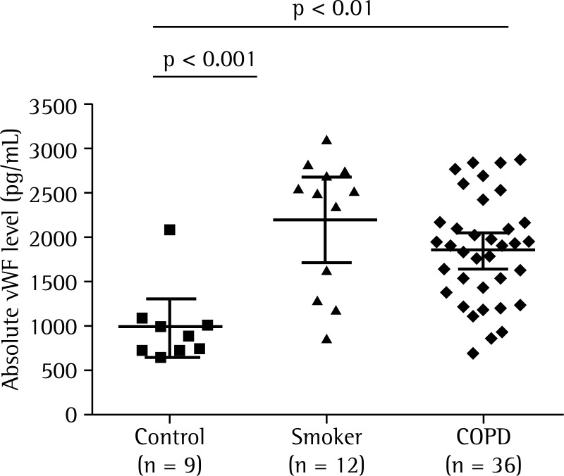
Objective: To compare the absolute serum von Willebrand factor (vWF) levels and relative serum vWF activity in patients with clinically stable COPD, smokers without airway obstruction, and healthy never-smokers.
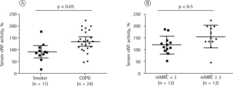
Methods: The study included 57 subjects, in three groups: COPD (n = 36); smoker (n = 12); and control (n = 9). During the selection phase, all participants underwent chest X-rays, spirometry, and blood testing. Absolute serum vWF levels and relative serum vWF activity were obtained by turbidimetry and ELISA, respectively. The modified Medical Research Council scale (cut-off score = 2) was used in order to classify COPD patients as symptomatic or mildly symptomatic/asymptomatic.
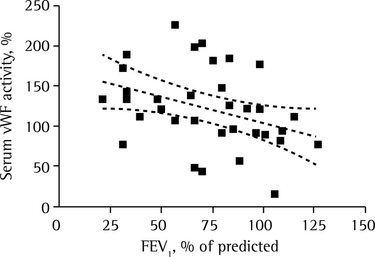
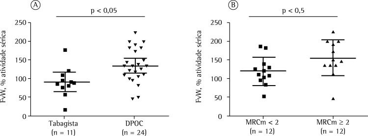
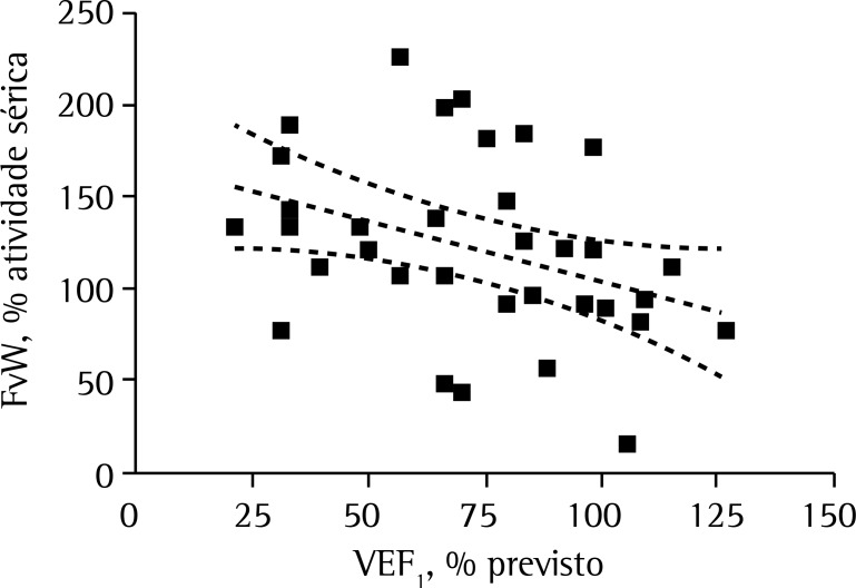
Results: Absolute vWF levels were significantly lower in the control group than in the smoker and COPD groups: 989 ± 436 pg/mL vs. 2,220 ± 746 pg/mL (p < 0.001) and 1,865 ± 592 pg/mL (p < 0.01). Relative serum vWF activity was significantly higher in the COPD group than in the smoker group (136.7 ± 46.0% vs. 92.8 ± 34.0%; p < 0.05), as well as being significantly higher in the symptomatic COPD subgroup than in the mildly symptomatic/asymptomatic COPD subgroup (154 ± 48% vs. 119 ± 8%; p < 0.05). In all three groups, there was a negative correlation between FEV1 (% of predicted) and relative serum vWF activity (r2 = -0.13; p = 0.009).
Conclusions: Our results suggest that increases in vWF levels and activity contribute to the persistence of systemic inflammation, as well as increasing cardiovascular risk, in COPD patients.
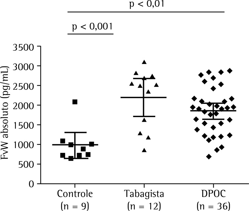
OBJETIVO:: Comparar os níveis séricos absolutos e a atividade sérica em percentual do fator de von Willebrand (FvW) em pacientes com DPOC clinicamente estáveis, tabagistas sem obstrução das vias aéreas e em indivíduos saudáveis que nunca fumaram.
MÉTODOS:: Foram incluídos no estudo 57 indivíduos, em três grupos: DPOC (n = 36), tabagista (n = 12) e controle (n = 9). Todos os participantes realizaram radiografia do tórax, espirometria e exame de sangue durante a fase de seleção. Os níveis séricos absolutos e a atividade sérica em percentual do FvW foram obtidos por turbidimetria e ELISA, respectivamente. A escala Medical Research Council modificada foi utilizada para classificar pacientes como sintomáticos ou assintomáticos/pouco sintomáticos no grupo DPOC (ponto de corte = 2).
RESULTADOS:: Os níveis absolutos do FvW no grupo controle foram significativamente menores que os nos grupos tabagista e DPOC: 989 ± 436 pg/mL vs. 2.220 ± 746 pg/mL (p < 0,001) e 1.865 ± 592 pg/mL (p < 0,01). Os valores em percentual de atividade do FvW no grupo DPOC foram significativamente maiores que no grupo tabagista (136,7 ± 46,0% vs. 92,8 ± 34,0%; p < 0,05), assim como foram significativamente maiores no subgrupo DPOC sintomático que no subgrupo DPOC assintomático/pouco sintomático (154 ± 48% vs. 119 ± 8%; p < 0,05). Houve uma correlação negativa entre o VEF1 (% do previsto) e os níveis em percentual de atividade do FvW nos três grupos (r2 = −0,13; p = 0,009).
CONCLUSÕES:: Nossos resultados sugerem que aumentos nos níveis de FvW e de sua atividade contribuem para a manutenção da inflamação sistêmica e o aumento do risco cardiovascular em pacientes com DPOC.
Figures
Similar articles
-
Increased von Willebrand Factor Processing in COPD, Reflecting Lung Epithelium Damage, Is Associated with Emphysema, Exacerbations and Elevated Mortality Risk.Int J Chron Obstruct Pulmon Dis. 2020 Mar 9;15:543-552. doi: 10.2147/COPD.S235673. eCollection 2020. Int J Chron Obstruct Pulmon Dis. 2020. PMID: 32210548 Free PMC article.
-
Biomarkers of Prothrombotic State and Risk Assessment of Exacerbations in Patients with Chronic Obstructive Pulmonary Disease.Int J Chron Obstruct Pulmon Dis. 2024 Oct 11;19:2273-2283. doi: 10.2147/COPD.S466563. eCollection 2024. Int J Chron Obstruct Pulmon Dis. 2024. PMID: 39416877 Free PMC article.
-
Microalbuminuria, von Willebrand factor and fibrinogen levels as markers of the severity in COPD exacerbation.J Thromb Thrombolysis. 2008 Oct;26(2):97-102. doi: 10.1007/s11239-007-0073-1. Epub 2007 Jul 10. J Thromb Thrombolysis. 2008. PMID: 17622488
-
Plasma levels of von Willebrand factor in type 2 diabetes patients with and without cardiovascular diseases: A meta-analysis.Diabetes Metab Res Rev. 2020 Jan;36(1):e3193. doi: 10.1002/dmrr.3193. Epub 2019 Jul 7. Diabetes Metab Res Rev. 2020. PMID: 31145835 Review.
-
von Willebrand factor and its relevance to cardiovascular disorders.Br Heart J. 1995 Dec;74(6):580-3. doi: 10.1136/hrt.74.6.580. Br Heart J. 1995. PMID: 8541159 Free PMC article. Review.
Cited by
-
Increased von Willebrand Factor Processing in COPD, Reflecting Lung Epithelium Damage, Is Associated with Emphysema, Exacerbations and Elevated Mortality Risk.Int J Chron Obstruct Pulmon Dis. 2020 Mar 9;15:543-552. doi: 10.2147/COPD.S235673. eCollection 2020. Int J Chron Obstruct Pulmon Dis. 2020. PMID: 32210548 Free PMC article.
-
Assessment of incidence of cerebral vascular diseases and prediction of stroke risk in chronic obstructive pulmonary disease patients using multimodal biomarkers.Clin Respir J. 2023 Mar;17(3):211-228. doi: 10.1111/crj.13587. Epub 2023 Jan 25. Clin Respir J. 2023. PMID: 36696969 Free PMC article.
-
Biomarkers of Clot Activation and Degradation and Risk of Future Major Cardiovascular Events in Acute Exacerbation of COPD: A Cohort Sub-Study in a Randomized Trial Population.Biomedicines. 2022 Aug 19;10(8):2011. doi: 10.3390/biomedicines10082011. Biomedicines. 2022. PMID: 36009558 Free PMC article.
-
Biomarkers of Prothrombotic State and Risk Assessment of Exacerbations in Patients with Chronic Obstructive Pulmonary Disease.Int J Chron Obstruct Pulmon Dis. 2024 Oct 11;19:2273-2283. doi: 10.2147/COPD.S466563. eCollection 2024. Int J Chron Obstruct Pulmon Dis. 2024. PMID: 39416877 Free PMC article.
-
Platelet activation and COPD-related clinical and imaging characteristics: The Multi-Ethnic Study of Atherosclerosis (MESA) COPD Study.Respir Med. 2025 May;241:108058. doi: 10.1016/j.rmed.2025.108058. Epub 2025 Mar 25. Respir Med. 2025. PMID: 40147570
References
-
- Macnee W. Pathogenesis of chronic obstructive pulmonary disease. Clin Chest Med. 2007;28(3):479–513. http://dx.doi.org/10.1016/j.ccm.2007.06.008 - DOI - PubMed
-
- Gan WQ, Man SF, Senthilselvan A, Sin DD. Association between chronic obstructive pulmonary disease and systemic inflammation: a systematic review and a meta-analysis. Thorax. 2004;59(7):574–580. http://dx.doi.org/10.1136/thx.2003.019588 - DOI - PMC - PubMed
-
- Ito K, Barnes PJ. COPD as a disease of accelerated lung aging. Chest. 2009;135(1):173–180. http://dx.doi.org/10.1378/chest.08-1419 - DOI - PubMed
-
- Warnier MJ, Rutten FH, Numans ME, Kors JA, Tan HL, de Boer A, et al. Electrocardiographic characteristics of patients with chronic obstructive pulmonary disease. COPD. 2013;10(1):62–71. http://dx.doi.org/10.3109/15412555.2012.727918 - DOI - PubMed
-
- Topsakal R, Kalay N, Ozdogru I, Cetinkaya Y, Oymak S, Kaya MG, et al. Effects of chronic obstructive pulmonary disease on coronary atherosclerosis. Heart Vessels. 2009;24(3):164–168. http://dx.doi.org/10.1007/s00380-008-1103-4 - DOI - PubMed
Publication types
MeSH terms
Substances
LinkOut - more resources
Full Text Sources
Other Literature Sources
Medical
Miscellaneous
