. 2014 Sep 11;9(9):e105984.
doi: 10.1371/journal.pone.0105984. eCollection 2014.

Cleavage specificity analysis of six type II transmembrane serine proteases (TTSPs) using PICS with proteome-derived peptide libraries

Affiliations

Cleavage specificity analysis of six type II transmembrane serine proteases (TTSPs) using PICS with proteome-derived peptide libraries

Olivier Barré et al. PLoS One. .

Abstract

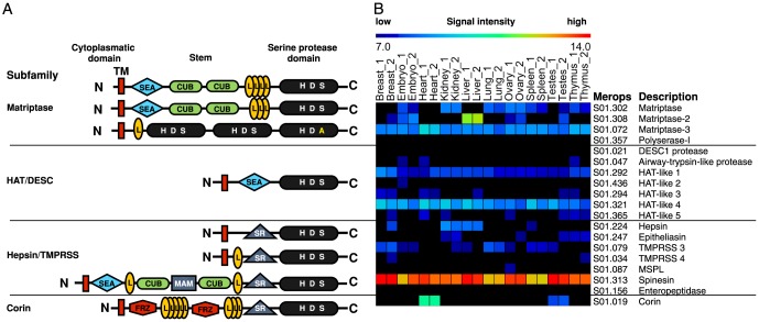
Background: Type II transmembrane serine proteases (TTSPs) are a family of cell membrane tethered serine proteases with unclear roles as their cleavage site specificities and substrate degradomes have not been fully elucidated. Indeed just 52 cleavage sites are annotated in MEROPS, the database of proteases, their substrates and inhibitors.

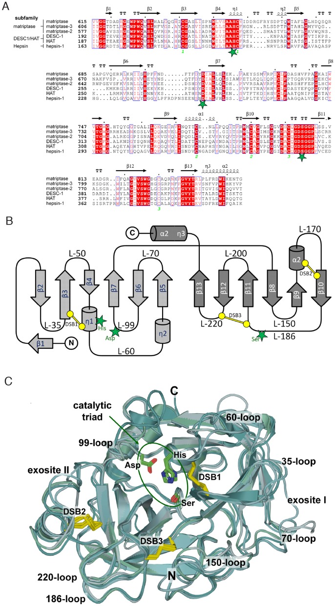
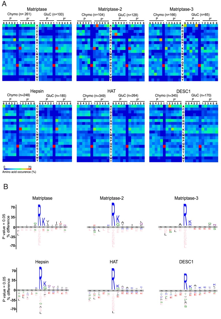
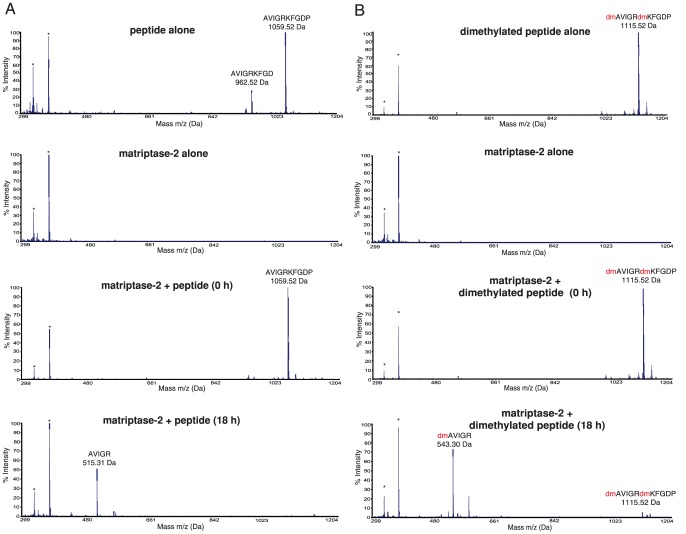
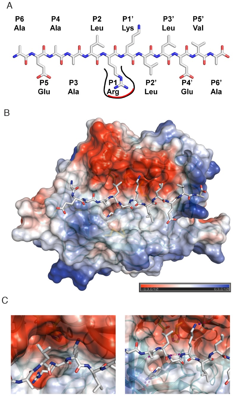
Methodology/principal finding: To profile the active site specificities of the TTSPs, we applied Proteomic Identification of protease Cleavage Sites (PICS). Human proteome-derived database searchable peptide libraries were assayed with six human TTSPs (matriptase, matriptase-2, matriptase-3, HAT, DESC and hepsin) to simultaneously determine sequence preferences on the N-terminal non-prime (P) and C-terminal prime (P') sides of the scissile bond. Prime-side cleavage products were isolated following biotinylation and identified by tandem mass spectrometry. The corresponding non-prime side sequences were derived from human proteome databases using bioinformatics. Sequencing of 2,405 individual cleaved peptides allowed for the development of the family consensus protease cleavage site specificity revealing a strong specificity for arginine in the P1 position and surprisingly a lysine in P1' position. TTSP cleavage between R↓K was confirmed using synthetic peptides. By parsing through known substrates and known structures of TTSP catalytic domains, and by modeling the remainder, structural explanations for this strong specificity were derived.

Conclusions: Degradomics analysis of 2,405 cleavage sites revealed a similar and characteristic TTSP family specificity at the P1 and P1' positions for arginine and lysine in unfolded peptides. The prime side is important for cleavage specificity, thus making these proteases unusual within the tryptic-enzyme class that generally has overriding non-prime side specificity.


Conflict of interest statement

Competing Interests: The authors have declared that no competing interests exist.

Figures

Figure 1
Figure 1. A. Schematic representation of the protein structures and arrangements of the four TTSP subfamilies: HAT/DESC, hepsin/TMPRSS, matriptase and corin.
All TTSPs contain a N-terminal transmembrane signal anchor domain (TM) and a C-terminal serine protease domain (H D S). In case of polyserase-1 one of the 3 catalytic domains is inactive (H D A). The stem region of the TTSPs contains 1–6 different domains: the sea urchin sperm protein/enteropeptidase/agrin domain (SEA), group A scavenger receptor domain (SR), low-density lipoprotein receptor class A domain (L), Cls/CLr, urchin embryonic growth factor, bone morphogenetic protein-1 domain (CUB) and Corin contains two frizzled (FRIZ) domain. B. Murine CLIP-CHIP RNA expression profile and its distribution of 19 members of the TTSP family in 10 murine tissues in duplicates according to their average signal intensity.
Figure 2
Figure 2. A. Multiple sequence alignment of the catalytic domains of six TTSPs was performed with Clustal Omega and displayed using ESPript 2.2 (http://esprit.ibcp.fr/ESPript/cgi-bin/ESPript.cgi .
Green stars indicate the residues of the catalytic triad; conserved residues are shaded in red. Blue boxes indicate amino acid with similar physico-chemical properties. Secondary structure elements are represented with flat arrows for β sheets and helices for α- and 310- (η) helices. Three conserved disulfide bridges are indicated in orange numerals below the alignment. Sequence numbering is according to the respective UniProt entries. B. Topology diagram of a TTSP catalytic domain. Residues of the catalytic triad are indicated as green stars, disulfide bonds (DSB) are shown in yellow. Loops (L-) are labeled corresponding to the thrombin nomenclature suggested by Bode et al. . C. Superposition of the catalytic domains of matriptase, matriptase-2, matriptase-3, hepsin, DESC1 and HAT, including both known and modeled structures, in traditional serine protease standard orientation . Catalytic triad and conserved disulfide bridges are shown in ball and stick representation. Important loops and exosites are indicated and labeled.
Figure 3
Figure 3. A. Matriptase, matriptase-2, matriptase-3, hepsin, DESC1, an HAT cleavage site specificities obtained by PICS using two different peptide libraries: GluC and chymotryptic.
For each class of peptide library, the amino acid occurrences in P6–P6′ are shown as heat maps. Above each heat map, the number of identified cleaved peptides is specified. B. Taking into account the relative natural abundances of amino acids, iceLogo representations of the different cleavage site specificities obtained from the GluC library are shown. Peptide sequences were aligned on the scissile bond between P1 and P1′. Statistically significant amino acid residue occurrences present (P<0.01) were plotted and amino acid heights are indicative for the degree of conservation at the indicated position. Residues that were completely absent in the identified terminal peptides are shown below in pink.
Figure 4
Figure 4
A. Upper panel. MALDI-TOF spectrum of the synthetic peptide AVIGRKFGDP. The sample contained a minor synthesis contaminant of AVIGRKFGD. The experimental determined [M+H]+ is 1059.52 Da (predicted m/z is 1059.22 Da). The sequence is presented by the spectra. The two asterix peaks represent the MALDI matrix peaks which are found in all the spectra in this m/z range. Second panel. MALDI-TOF spectrum of matriptase-2. Third panel. MALDI-TOF spectrum of the synthetic peptide added to matriptase-2 at 0 h. Lower panel. MALDI-TOF spectrum of the assay reaction products generated after incubation of the synthetic peptide with matriptase-2 for 18 h. The spectral peak at 1059.52 m/z disappeared and a new peak at 515.31 Da appeared corresponding to the cleavage product [AVIGR+H]+ (predicted m/z is 514.96 Da). B. Upper panel. MALDI-TOF spectrum of the dimethylated (dm) synthetic peptide (dm)AVIGR(dm)KFGDP. The experimental determined [M+H]+ is 1115.52 Da. The sequence is presented on the spectral peak. The two asterix peaks represent the MALDI matrix peaks and they can be found in all the spectra. Second panel. MALDI-TOF spectrum of matriptase-2. Third panel. MALDI-TOF spectrum of the reaction products after incubation of the dimethylated synthetic peptide with matriptase-2 added at 0 h. Lower panel. MALDI-TOF spectrum of the reaction products after incubation of the dimethylated synthetic peptide with matriptase-2 for 18 h. The peak at 1115.52 Da disappeared and a new peak at 543.30 Da appeared, corresponding to [dAVIGR+H]+. Red (dm) is dimethylation.
Figure 5
Figure 5. A Schematic representation of the dodecapeptide (AEAALR↓KLLEVA) used for active site docking.
The deep TTSP S1 subsite, accommodating the P1-Arg, is shown. B. Structural model of matriptase-2 with the peptide substrate (AEAALRKLLEVA) identified by PICS docked into the active site. Matriptase-2 was colored according to its surface charge distribution, and is shown in standard orientation with the modeled peptide occupying subsites S6 to S6′ (from left to right). C. Close-up views of the active site illustrating the deep and negatively charged S1 pocket (left) and key stabilizing interaction between the protease and the modeled peptide (right).

References

    1. Leytus SP, Loeb KR, Hagen FS, Kurachi K, Davie EW (1988) A novel trypsin-like serine protease (hepsin) with a putative transmembrane domain expressed by human liver and hepatoma cells. Biochemistry 27: 1067–1074. - PubMed
    1. Rodríguez D, Morrison CJ, Overall CM (2010) Matrix metalloproteinases: What do they not do? New substrates and biological roles identified by murine models and proteomics. Biochimica et Biophysica Acta (BBA) - Molecular Cell Research 1803: 39–54 10.1016/j.bbamcr.2009.09.015 - DOI - PubMed
    1. Overall CM, Blobel CP (2007) In search of partners: linking extracellular proteases to substrates. Nat Rev Mol Cell Biol 8: 245–257 10.1038/nrm2120 - DOI - PubMed
    1. Netzel-Arnett S, Hooper JD, Szabo R, Madison EL, Quigley JP, et al. (2003) Membrane anchored serine proteases: a rapidly expanding group of cell surface proteolytic enzymes with potential roles in cancer. Cancer Metastasis Rev 22: 237–258. - PubMed
    1. Szabo R, Bugge TH (2008) Type II transmembrane serine proteases in development and disease. Int J Biochem Cell Biol 40: 1297–1316 10.1016/j.biocel.2007.11.013 - DOI - PubMed

Publication types