. 2014 Sep 11;10(9):e1004366.
doi: 10.1371/journal.ppat.1004366. eCollection 2014 Sep.

Sialylation of prion protein controls the rate of prion amplification, the cross-species barrier, the ratio of PrPSc glycoform and prion infectivity

Affiliations

Sialylation of prion protein controls the rate of prion amplification, the cross-species barrier, the ratio of PrPSc glycoform and prion infectivity

Elizaveta Katorcha et al. PLoS Pathog. .

Abstract

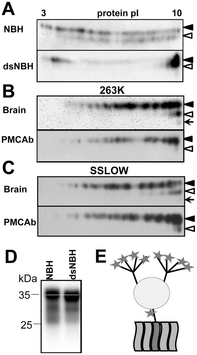
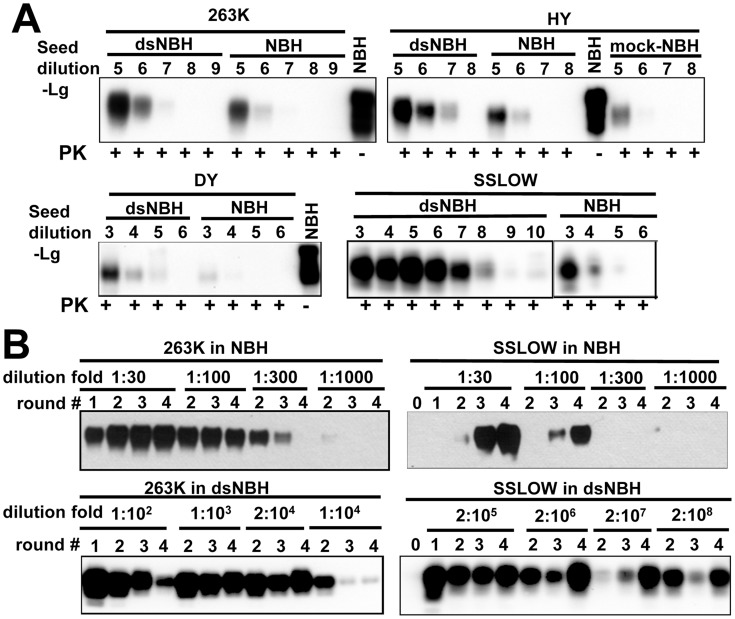
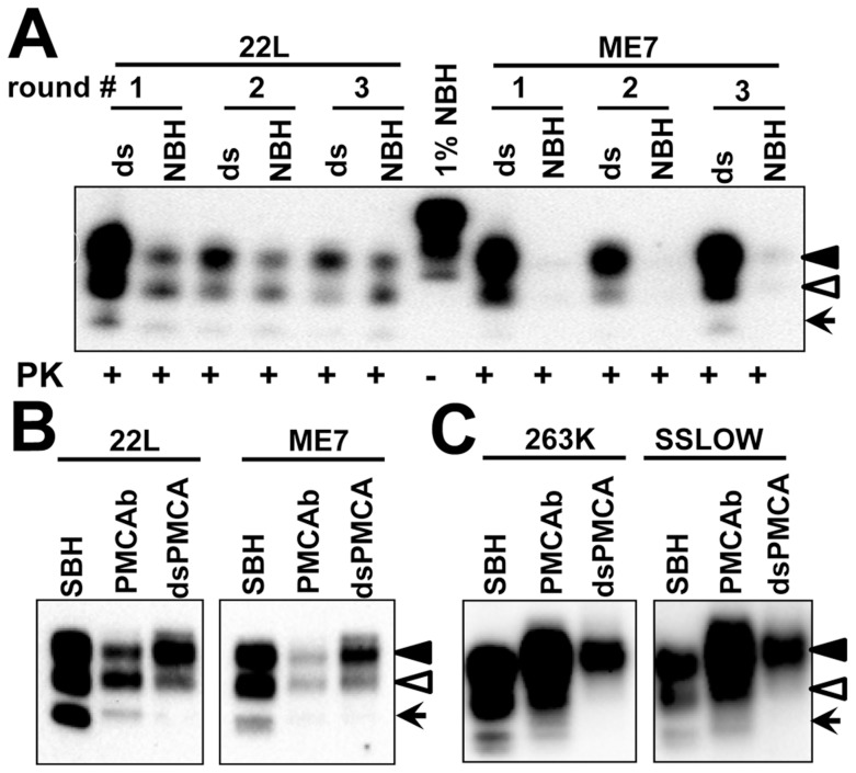
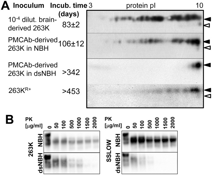
The central event underlying prion diseases involves conformational change of the cellular form of the prion protein (PrP(C)) into the disease-associated, transmissible form (PrP(Sc)). Pr(PC) is a sialoglycoprotein that contains two conserved N-glycosylation sites. Among the key parameters that control prion replication identified over the years are amino acid sequence of host PrP(C) and the strain-specific structure of PrPSc. The current work highlights the previously unappreciated role of sialylation of PrP(C) glycans in prion pathogenesis, including its role in controlling prion replication rate, infectivity, cross-species barrier and PrP(Sc) glycoform ratio. The current study demonstrates that undersialylated PrP(C) is selected during prion amplification in Protein Misfolding Cyclic Amplification (PMCAb) at the expense of oversialylated PrP(C). As a result, PMCAb-derived PrP(Sc) was less sialylated than brain-derived PrP(Sc). A decrease in PrPSc sialylation correlated with a drop in infectivity of PMCAb-derived material. Nevertheless, enzymatic de-sialylation of PrP(C) using sialidase was found to increase the rate of PrP(Sc) amplification in PMCAb from 10- to 10,000-fold in a strain-dependent manner. Moreover, de-sialylation of PrP(C) reduced or eliminated a species barrier of for prion amplification in PMCAb. These results suggest that the negative charge of sialic acid controls the energy barrier of homologous and heterologous prion replication. Surprisingly, the sialylation status of PrP(C) was also found to control PrP(Sc) glycoform ratio. A decrease in Pr(PC) sialylation levels resulted in a higher percentage of the diglycosylated glycoform in PrP(Sc). 2D analysis of charge distribution revealed that the sialylation status of brain-derived PrP(C) differed from that of spleen-derived PrP(C). Knocking out lysosomal sialidase Neu1 did not change the sialylation status of brain-derived PrP(C), suggesting that Neu1 is not responsible for desialylation of PrP(C). The current work highlights previously unappreciated role of PrP(C) sialylation in prion diseases and opens multiple new research directions, including development of new therapeutic approaches.


Conflict of interest statement

The authors have declared that no competing interests exist.

Figures

Figure 1
Figure 1. 2D analysis of PrPC and brain- and PMCAb-derived PrPSc.
A. 2D analysis of Syrian hamster NBH (top panel) and NBH treated with sialidase from A. ureafaciens (bottom gel). B, C. 2D analysis of brain- and PMCAb-derived 263K material (B), and brain- and PMCAb-derived SSLOW material (C). To produce PMCAb-derived material, brain-derived 263K or SSLOW was subjected to 24 serial rounds with 10-fold dilution between rounds. Black and white triangles mark diglycosylated and monoglycosylated glycoforms, respectively, whereas arrows mark the unglycosylated form. D. Migration of PrPC in 1D SDS-PAGE gel before and after treatment with sialidase. All blots were stained with 3F4 antibody. E. Schematic diagram of PrPC that illustrates location of sialic acid residues (stars) on N-linked glycans and GPI anchor. Each of the two glycans can carry up to four terminal sialic acid residues.
Figure 2
Figure 2. De-sialylation of PrPC increases the rate of PrPSc amplification in PMCAb.
A. 263K, HY, Drowsy or SSLOW scrapie brain materials were diluted 103–1010-fold into 10% NBH, dsNBH or mock treated NBH (NBH incubated with a buffer for enzymatic de-sialylation in the absence of sialidase) as indicated and subjected to a single PMCAb round. Undigested 10% NBH was used as reference. B. Analysis of PrPSc amplification fold. Scrapie brain materials were diluted 104-fold for 263K or 103-fold for SSLOW into 10% NBH or dsNBH as indicated and subjected to four serial PMCAb rounds. The material amplified in each round was diluted to a specified dilution fold into 10% NBH or dsNBH for the next PMCAb round as indicated. Unamplified seeds are shown as round 0. Prior to electrophoresis, samples were treated with PK. All blots were stained with 3F4 antibody.
Figure 3
Figure 3. De-sialylation of PrPC reduces species barrier in PMCAb.
A. 263K or HY brain materials were diluted 103-fold into 10% mouse dsNBH or NBH and subjected to ten serial PMCAb rounds with 5-fold dilution between rounds. Prior to electrophoresis, samples were treated with PK. Blots were stained with Ab3531 antibody. B. 22L or ME7 brain material was diluted 103-fold into 10% hamster dsNBH or NBH and subjected to ten serial PMCAb rounds with 5-fold dilution between rounds. Prior to electrophoresis samples were treated with PK. Blots were stained with 3F4 antibody. Black and white triangles mark diglycosylated and monoglycosylated forms, respectively. C. PK-resistance profile illustrating relative representation of di-, mono-, and unglycosylated glycoforms in 263K-seeded PMCAb products produced in three reactions with mouse NBH (bold solid lines) or dsNBH (dashed lines).
Figure 4
Figure 4. Influence of PrPC sialylation level on glycoform distribution in PMCAb-derived products.
A. 22L or ME7 brain material was diluted 5×10²- or 2×10³-fold, respectively, into 10% mouse dsNBH or NBH and subjected to three serial PMCAb rounds with 5-fold dilution between rounds. Prior to electrophoresis samples were treated with PK. Blot was stained with Ab3531 antibody. Undigested 1% mouse NBH was used as a reference. B. Western blot of 22L or ME7 scrapie brain material (SBH) or material produced in PMCAb using NBH or dsNBH (dsPMCAb), and stained with Ab3531 antibody. C. Western blot of 263K or SSLOW scrapie brain material (SBH) or material produced in PMCAb using NBH or dsNBH (dsPMCAb), and stained with 3F4 antibody. Black and white triangles mark di- and mono-glycosylated glycoforms, respectively, whereas arrows mark the unglycosylated form.
Figure 5
Figure 5. 2D analysis of brain- and spleen-derived PrPC.
2D analysis of Syrian hamster spleen (A) and brain homogenates (B). Diglycosylated and monoglycosylated full-length PrPC (FL) are marked by black and white triangles, respectively. Di-, mono- and unglycosylated C2 fragments (C2) in spleen or C1 fragments (C1) in brain are marked by bold, medium and thin arrows, respectively. PrPC, C1 or C2 dimers (D) are encircled. M stands for a marker lane: brain or spleen samples were diluted 10-fold and used as references for 2D gels. Blots were stained with SAF-84 antibody.
Figure 6
Figure 6. Correlation between PrPSc sialylation status and its infectivity.
A. The following material was analyzed by 2D and animal bioassay: 1% brain-derived 263K material, PMCAb-derived 263K (products of 24th PMCAb rounds conducted in NBH with 10-fold dilution between rounds), PMCAb-derived 263K produced in dsNBH (products of 7th PMCAb round conducted in dsNBH with 1000-fold dilution between rounds) and 263KR+ (263K brain material subjected to 12 serial PMCAb rounds in RNA-depleted NBH and then an additional 14 PMCAb rounds in NBH as described in [46]). Prior to inoculation, all PMCAb-derived materials were diluted 10-fold. 104-fold diluted 263K brain material was used for inoculating a reference group to match the amount of PK-resistant material in PMCAb-derived samples. Diglycosylated and monoglycosylated PrPC are marked by black and white triangles, respectively. B. Western blots of 263K- or SSLOW-seeded PMCAb-derived material produced using NBH or dsNBH and treated with increasing concentrations of PK as indicated. All blots were stained with 3F4 antibody.
Figure 7
Figure 7. Schematic diagrams illustrating that prion polymerization and PrPSc glycoform ratio are controlled by sialic acid residues of glycans.
A, B. Sialylation of glycans impedes prion amplification in PMCAb due to electrostatic repulsion between sialic acid residues on the PrPSc surface. Desialylation of a substrate facilitates prion polymerization in dsPMCAb. Due to strain-specific differences in PrPSc structures, PrPC desialylation speeds up polymerization of strains of synthetic origin (B) much stronger than strains of natural origin (A). C. Electrostatic repulsion between sialic acid residues limits percentage of diglycosylated glycoforms that can be accommodated in PrPSc. Desialylation of PrPC in dsPMCAb changes the ratio of glycoforms in PrPSc in favor of diglycosylated glycoforms. PrPC monomers are depicted as blue circles; glycosyls are shown as yellow circles where presence of terminal sialic acid residues is shown as a red glow. Electrostatic repulsion is represented with lightning bolts.

Similar articles

Cited by

References

    1. Prusiner SB (1998) Prions. Proc Natl Acad Sci U S A 95: 13363–13383. - PMC - PubMed
    1. Cohen FE, Prusiner SB (1998) Pathologic conformations of prion proteins. Annu Rev Biochem 67: 793–819. - PubMed
    1. Stahl N, Baldwin MA, Hecker R, Pan KM, Burlingame AL, et al. (1992) Glycosylinositol phospholipid anchors of the scrapie and cellular prion proteins contain sialic acid. Biochemistry 31: 5043–5053. - PubMed
    1. Stahl N, Borchelt DR, Hsiao K, Prusiner SB (1987) Scrapie prion protein contains a phosphatidylinositol glycolipid. Cell 51: 229–240. - PubMed
    1. Turk E, Teplow DB, Hood LE, Prusiner SB (1988) Purification and properties of the cellular and scrapie hamster prion proteins. Eur J Biochem 176: 21–30. - PubMed

Publication types

MeSH terms