. 2014 Sep 10;16(3):314-327.
doi: 10.1016/j.chom.2014.07.015.

The interferon signaling antagonist function of yellow fever virus NS5 protein is activated by type I interferon

Affiliations

The interferon signaling antagonist function of yellow fever virus NS5 protein is activated by type I interferon

Maudry Laurent-Rolle et al. Cell Host Microbe. .

Abstract

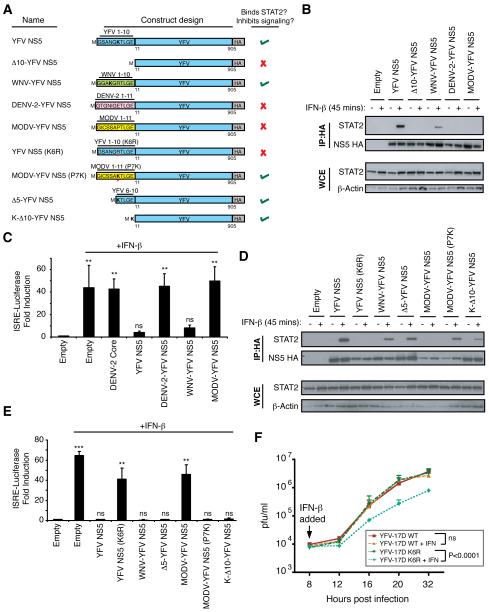
To successfully establish infection, flaviviruses have to overcome the antiviral state induced by type I interferon (IFN-I). The nonstructural NS5 proteins of several flaviviruses antagonize IFN-I signaling. Here we show that yellow fever virus (YFV) inhibits IFN-I signaling through a unique mechanism that involves binding of YFV NS5 to the IFN-activated transcription factor STAT2 only in cells that have been stimulated with IFN-I. This NS5-STAT2 interaction requires IFN-I-induced tyrosine phosphorylation of STAT1 and the K63-linked polyubiquitination at a lysine in the N-terminal region of YFV NS5. We identified TRIM23 as the E3 ligase that interacts with and polyubiquitinates YFV NS5 to promote its binding to STAT2 and trigger IFN-I signaling inhibition. Our results demonstrate the importance of YFV NS5 in overcoming the antiviral action of IFN-I and offer a unique example of a viral protein that is activated by the same host pathway that it inhibits.


Figures

Figure 1
Figure 1. YFV inhibits IFN-I but not IFN-II signaling
293T cells were transfected with (A) an IFN-I-inducible firefly-luciferase (f-luc) reporter plasmid (ISRE-luciferase) or (B) an IFN-II-inducible f-luc reporter (GAS-luciferase) along with a constitutively expressing Renilla luciferase (R-Luc) gene plasmid. Cells were infected with DENV-2 or YFV-17D viruses (MOI=5) for 24 hrs and treated with IFN-β or IFN-γ (1000 U/ml) for 16 hrs prior to assaying for luciferase activity. Fold induction of f-luc was normalized to R-luc. The bars represent the mean fold induction of 3 independent experiments compared to untreated, mock-infected controls. (C) Vero cells were mock infected or infected with YFV-17D virus (MOI=5). At 24 hrs post infection (hpi), cells were mock treated or treated with the indicated amounts of IFN-β or IFN-γ for 16 hrs then infected with NDV-GFP. NDV-GFP replication was monitored by fluorescence microscopy at 14 hpi. (D, E) Confocal microscopy images showing subcellular localization of the indicated protein in mock-infected or YFV-17D-infected (MOI=1) Vero cells before or after treatment with IFN-β for 30 minutes. (F) Intracellular fractionation of mock infected or YFV-17D-infected HeLa cells (MOI=10). The proteins in the cytoplasmic and nuclear fractions were detected by western blotting with the indicated antibodies. (G) Vero cells were mock-infected or infected with DENV-2 or YFV-17D viruses for 24 hours (MOI=10), then either mock-stimulated or stimulated with IFN-β for 8 hours. Cell extracts were analyzed by EMSA with ISRE elements derived from theOas1b gene. Statistical analyses were conducted using the unpaired t test in Prism 4 for Macintosh (GraphPad Software, USA). ns = no significance, where p>0.05; *** = p<0.0001.
Figure 2
Figure 2. YFV NS5 inhibits IFN-I signaling and interacts with STAT2 in IFN-I- and IFN-III-treated cells
(A) 293T cells were co-transfected with a plasmid encoding an ISRE-54-CATGFP reporter and with a constitutively expressing firefly-luciferase (f-luc) plasmid plus an empty vector or viral protein encoding plasmids. At 24 hrs post transfection, cells were treated with IFN-β for 16 hrs prior to assaying for CAT activity. Induction of CAT activity was normalized to fluc activity. The lower panel of (A) is a western blot showing the relative expression of each transfected viral protein. (B) 293T cells were co-transfected with an IFN-I-inducible f-luc reporter plasmid (pISRE-luc) along with a constitutively expressing Renilla luciferase (R-luc) gene plasmid and empty vector or YFV-17D and YFV-Asibi NS5 expression plasmids. At 24 hrs post transfection, cells were treated with IFN-β for 16 hrs prior to assaying for luciferase activities. Fold induction of f-luc was normalized to R-luc. The bars represent the mean fold induction of 3 independent experiments compared to untreated empty vector controls. The lower panel of (B) is a western blot (WB) showing the relative expression of each transfected viral protein. (C) 293T cells were transfected with the indicated NS5 expression plasmids. At 24 hrs, cells were mock stimulated or stimulated with IFN-β for 45 minutes. Cells were lysed and immunoprecipitation was performed followed by WB with the indicated antibodies. (D) Same experiment as in (C) with cells mock-stimulated or stimulated with 1000 U/ml of IFN-β, IFN-γ or IFN-λ for 45 minutes prior to lysis. Abbreviations: WCE = whole cell extract; CAT = chloramphenicol acetyl transferase.
Figure 3
Figure 3. Neither IFN-I-mediated phosphorylation nor nuclear translocation of STAT2 are required for STAT2 interaction with YFV NS5
(A) Confocal microscopy images showing subcellular localization of the indicated proteins in Vero cells before or after treatment with IFN-β for 45 minutes. (B) U6A cells were transfected with the indicated plasmids. At 24 hours, cells were mock stimulated or stimulated with IFN-β for 45 minutes. Cells were lysed and immunoprecipitation was performed against the HA epitope for 2 hrs using anti-HA beads. The beads were then washed three times with lysis buffer. 2fTGH cells were mock stimulated or stimulated with IFN-β then lysed and added to the NS5-bound anti-HA beads for 2 additional hrs. The protein was eluted from washed beads then used for western blot.
Figure 4
Figure 4. A single lysine residue at position 6 of YFV NS5 is critical for IFN-I antagonist function
(A) Diagram of the YFV NS5 expression constructs used in Figure 4. (B and D) 293T cells were transfected with the indicated plasmids. At 24 hours post transfection, cells were mock stimulated or stimulated with IFN-β for 45 minutes. Cells were lysed and immunoprecipitation was performed followed by western blot analysis with the indicated antibodies. (C and E) 293T cells were co-transfected with an IFN-β-inducible firefly-luciferase (fluc) reporter plasmid (pISRE-luc) along with a constitutively expressing R-luc plasmid plus empty vector or plasmids encoding the indicated viral proteins. At 24 hrs, cells were treated with IFN-β for 16 hrs prior to assaying for luciferase activities. Fold induction of f-luc activity was normalized to R-luc activity. The bars represent the mean fold induction of 3 independent experiments compared to the untreated, empty vector controls. (F) Vero cells were infected with either YFV-17D WT or YFV-17D K6R at an MOI of 10. At 8 hpi, the cells were either mock-treated or treated with 100 U/ml IFN-β. Virus was harvested at the indicated time points and viral titers were quantified by plaque assay on BHK-21 cells. Each point on the graph represents the mean of 4 independent experiments. Statistical analyses were conducted using the unpaired t test in Prism 4 (GraphPad Software, USA). ns = no significance, where p>0.05; *** = p<0.0001; ** = p<0.001.
Figure 5
Figure 5. K63-linked ubiquitination is required for binding of YFV NS5 to STAT2
(A) 293T cells were transfected with various NS5 mutants. At 24 hrs post transfection, cells were mock stimulated or stimulated with IFN-β for 45 minutes. Cells were lysed and immunoprecipitation was performed followed by western blot analysis (WB) with the indicated antibodies. (B) Same as (A) but with more stringent conditions. (C) U6A cells were transfected with HA-tagged YFV NS5. At 24 hrs, cells were mock stimulated or stimulated with IFN-β. Cells were lysed and immunoprecipitation was performed against the HA epitope for 2 hrs using HA beads. Beads were washed three times with lysis buffer followed by incubation with reaction buffer or OTU. 2fTGH cells were mock stimulated or stimulated with 1000 U/ml IFN- β. Cells were lysed and were added to the HA beads for 2 hrs. Beads were then washed followed by WB with the indicated antibodies. (D) Diagram of the YFV NS5 constructs used in 5E and 5F. (E) Same experiment as in (A) with the indicated constructs. (F) 293T cells were co-transfected with an IFN-α/β-inducible firefly-luciferase (f-luc) reporter plasmid (pISRE-luc) along with a constitutively expressing Renilla luciferase (R-luc) gene plasmid plus empty vector or plasmids encoding the indicated viral proteins. At 24 hrs, cells were treated with IFN-β for 16 hrs prior to assaying for luciferase activities. Fold induction of f-luc activity was normalized to R-luc activity. The bars represent the mean fold induction of 3 independent experiments compared to the untreated empty vector controls. (G) 293T cells were transfected with FLAG-tagged wild-type YFV NS5 and various HA-tagged ubiquitin mutants. At 24 hours, cells were mock stimulated or stimulated with IFN-β for 45 minutes. Cells were lysed and immunoprecipitation was performed against the HA epitope followed by WB with the indicated antibodies. Statistical analyses were conducted using the unpaired t test in Prism 4 (GraphPad Software, USA). ns = no significance, where p>0.05; *** = p<0.0001.
Figure 6
Figure 6. TRIM23 ubiquitinates YFV NS5
(A) Confocal microscopy images showing subcellular localization of the indicated proteins in HeLa cells before or after treatment with IFN-β for 30 minutes. (B) 293T cells were transfected with YFV NS5. At 24 hours, cells were mock stimulated or stimulated with IFN-β then lysed followed by immunoprecipitation. Western blot analysis (WB) was done with the indicated antibodies. (C) Ubiquitination assay conducted in 293T cells with increasing amounts of TRIM23. Cells were transfected with FLAG-tagged wild-type YFV NS5 or YFV NS5 K6R with increasing amounts of HA-tagged TRIM23. Lysis and immunoprecipitation against the FLAG epitope was followed by western blot analysis with the indicated antibodies. (D) siRNA-expressing A549 cells were infected with YFV-17D WT at an MOI of 10. At 8 hpi, the cells were mock-treated or treated with 100 U/ml IFN-β. Virus was harvested at 24 hours and viral RNA, TRIM23 and MxA RNA quantified by qPCR. Viral titers were quantified by plaque assay on BHK-21 cells. (E) siRNA-expressing 293T cells were transfected with empty plasmid or YFV NS5 and mock treated or treated with IFN-I for 45 minutes. Lysis and immunoprecipitation were followed by WB with the indicated antibodies. Statistical analyses were conducted using the unpaired t test in Prism 4 (GraphPad Software, USA). ns = no significance, where p>0.05; *** = p<0.0001; ** = p<0.001.
Figure 7
Figure 7. STAT1 is necessary for IFN-I induced YFV NS5-STAT2 interaction
(A) 293T cells were transfected with the indicated constructs. Cells were mock stimulated or stimulated with IFN-β for 45 minutes at 24 hours post transfection. Cell lysates were immunoprecipitated followed by western blot analysis with the indicated antibodies. (B) Same experiment as in (A) done in U3A cells. In the second panel, U3A cells were complemented with wild-type STAT1 and the same experiment was repeated. (C) U3A cells were complemented with wild-type STAT1 or mutant STAT1 Y701F and the experimental procedure in (B) was repeated.

Comment in

References

    1. Aguirre S, Maestre AM, Pagni S, Patel JR, Savage T, Gutman D, Maringer K, Bernal-Rubio D, Shabman RS, Simon V, et al. DENV inhibits type I IFN production in infected cells by cleaving human STING. PLoS pathogens. 2012;8:e1002934. - PMC - PubMed
    1. Ank N, West H, Bartholdy C, Eriksson K, Thomsen AR, Paludan SR. Lambda interferon (IFN-lambda), a type III IFN, is induced by viruses and IFNs and displays potent antiviral activity against select virus infections in vivo. Journal of virology. 2006;80:4501–4509. - PMC - PubMed
    1. Arimoto K, Funami K, Saeki Y, Tanaka K, Okawa K, Takeuchi O, Akira S, Murakami Y, Shimotohno K. Polyubiquitin conjugation to NEMO by triparite motif protein 23 (TRIM23) is critical in antiviral defense. Proceedings of the National Academy of Sciences of the United States of America. 2010;107:15856–15861. - PMC - PubMed
    1. Ashour J, Laurent-Rolle M, Shi PY, Garcia-Sastre A. NS5 of dengue virus mediates STAT2 binding and degradation. Journal of virology. 2009;83:5408–5418. - PMC - PubMed
    1. Ashour J, Morrison J, Laurent-Rolle M, Belicha-Villanueva A, Plumlee CR, Bernal-Rubio D, Williams KL, Harris E, Fernandez-Sesma A, Schindler C, et al. Mouse STAT2 restricts early dengue virus replication. Cell host & microbe. 2010;8:410–421. - PMC - PubMed

Publication types

MeSH terms