Review
. 2014 Nov;121(5):1099-121.
doi: 10.1097/ALN.0000000000000446.

Cell-based therapy for acute organ injury: preclinical evidence and ongoing clinical trials using mesenchymal stem cells

Affiliations
Review

Cell-based therapy for acute organ injury: preclinical evidence and ongoing clinical trials using mesenchymal stem cells

Antoine Monsel et al. Anesthesiology. 2014 Nov.

Abstract

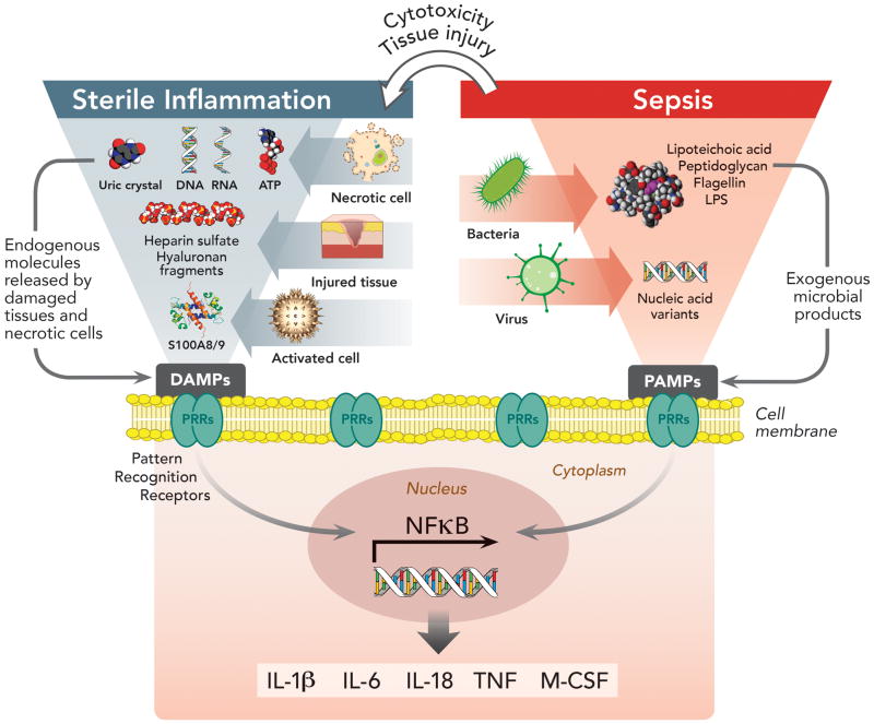
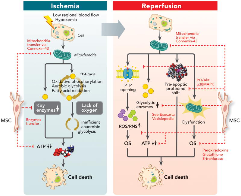
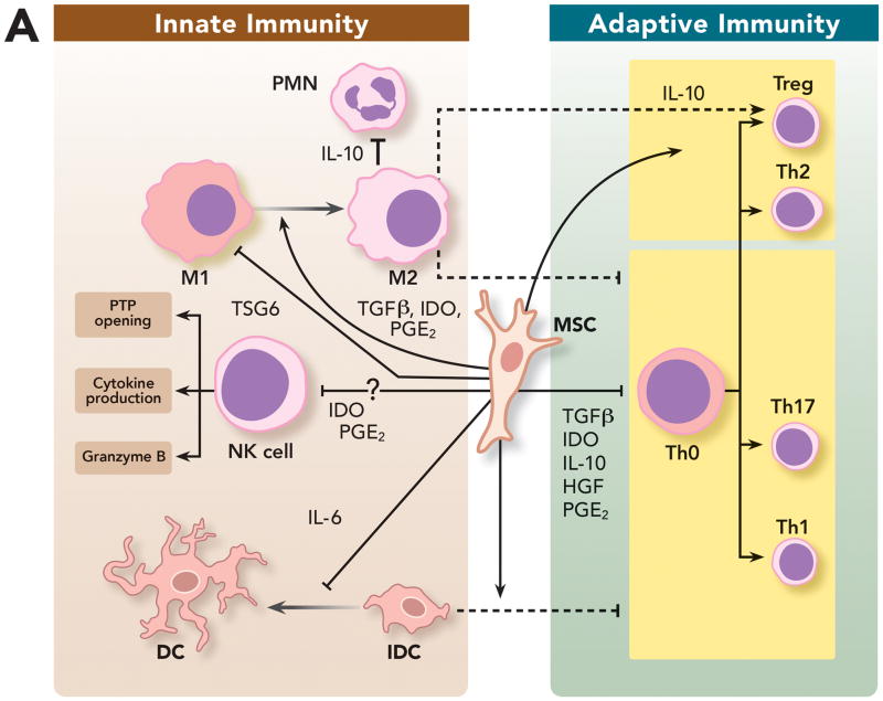
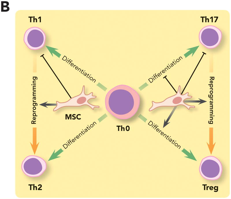
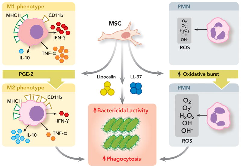
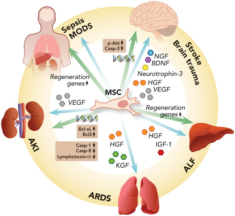
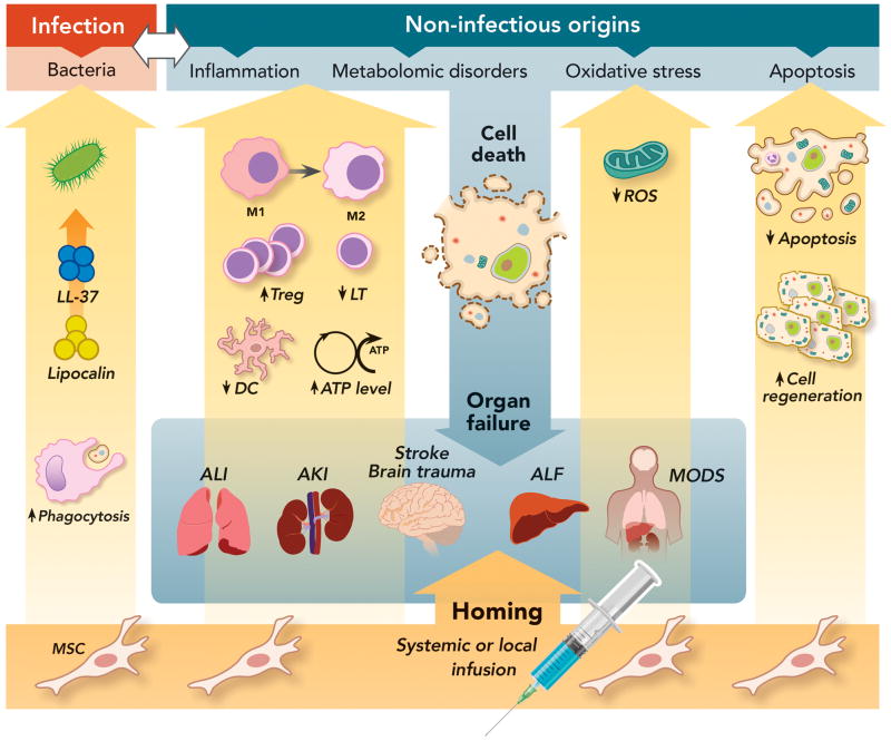
Critically ill patients often suffer from multiple organ failures involving lung, kidney, liver, or brain. Genomic, proteomic, and metabolomic approaches highlight common injury mechanisms leading to acute organ failure. This underlines the need to focus on therapeutic strategies affecting multiple injury pathways. The use of adult stem cells such as mesenchymal stem or stromal cells (MSC) may represent a promising new therapeutic approach as increasing evidence shows that MSC can exert protective effects following injury through the release of promitotic, antiapoptotic, antiinflammatory, and immunomodulatory soluble factors. Furthermore, they can mitigate metabolomic and oxidative stress imbalance. In this work, the authors review the biological capabilities of MSC and the results of clinical trials using MSC as therapy in acute organ injuries. Although preliminary results are encouraging, more studies concerning safety and efficacy of MSC therapy are needed to determine their optimal clinical use. (ANESTHESIOLOGY 2014; 121:1099-121).


Conflict of interest statement

The authors declare no competing interests.

Figures

Figure 1
Figure 1. Pattern Recognition Receptors in Immunity and Their Involvement in Sterile and Sepsis-Related Inflammation
Pattern recognition receptors (PRRs) expressed by antigen presenting cells (dendritic cells, monocytes, macrophages) constitute the first interaction between the extra-cellular environment and innate immunity. They are proteins, which include membrane-bound and cytoplasmic receptors, that bind either pathogen-associated molecular patterns (PAMPs) or damage-associated molecular patterns (DAMPs) derived from exogenous microorganisms (i.e. sepsis from infection) or endogenous molecules (i.e. sterile inflammation). Interaction of PRRs with PAMPs/DAMPs induces nuclear factor-kappa B signaling pathways, resulting in the secretion of pro-inflammatory cytokines and co-stimulatory molecules. In sepsis, the initial immune response triggered by PAMPs/PRRs interaction can lead to tissue damage and the release of DAMPs, which may act synergistically with PAMPs to enhance inflammation. Nevertheless, even without microorganism involvement, DAMPs released from dead or dying cells in response to injury or stress, are able to induce similar pro-inflammatory cytokine production from tissues, driving “sterile inflammation.” ATP = adenosine triphosphate; DAMPs = damage-associated molecular patterns; IL-1β = interleukin-1 beta; IL-6 = interleukin 6; IL-18 = interleukin 18; LPS = lipopolysaccharide; M-CSF = macrophage colony-stimulating factor; NF-κB = nuclear factor kappa-light-chain-enhancer of activated B cells; PAMPs = pathogen-associated molecular patterns; PRRs = pattern recognition receptors; S100A8/9 = (also known as calgranulins A and B, or MRP8 and MRP14 respectively) are members of the S100 multigene subfamily of cytoplasmic EF-hand Ca2+-binding proteins which are endogenous activators of Toll-like receptor 4; TNF = tumor necrosis factor.
Figure 2
Figure 2. Impact of Mesenchymal Stem Cells on Ischemia-Reperfusion Injury Pathways
Ischemia is a significant cause of acute organ injury that results from a decrease in regional oxygen delivery (such as low blood flow or hypoxemia), leading to inefficient anaerobic glycolysis as the major source of ATP production and ATP deficit. However, much of the tissue damage occurs during the reperfusion phase, leading to mitochondrial permeability transition pore opening, pro-glycolytic enzyme depletion, pro-apoptotic proteome shift and mitochondrial dysfunction inducing oxidative stress. MSC can decrease ischemia-reperfusion induced injury by: (1) Restoring ATP levels by possibly mitochondrial transfer through connexin-43 channels and replenishing depleted glycolytic enzymes; (2) Decreasing reactive oxygen species/reactive nitrogen species generated during oxidative stress by either preventing their release, circumventing the depletion of key enzymes or by transferring reactive oxygen species scavengers (such as peroxiredoxins and glutathione S-transferase) into injured cells; (3) And restoring proteomic alterations by activating pro-survival phosphatidylinositide 3-kinases/protein kinase B pathway via cluster of differentiation 73 or inhibiting p38 MAPK-caspase 3 pathway. ATP = adenosine triphosphate; CD73 = cluster of differentiation 73; MAPK = mitogen-activated protein kinases; MSC = mesenchymal stem cell; OS = oxidative stress; PI3/Akt = phosphatidylinositide 3-kinases/protein kinase B; PTP = permeability transition pore; ROS = reactive oxygen species; RNS = reactive nitrogen species; TCA = tricarboxylic acid cycle.
Figure 3
Figure 3. Immunomodulatory Properties of Mesenchymal Stem Cells on Innate and Adaptive Immunity
(A) MSC can modulate innate and adaptive immune cells by: (1) Promoting repolarization of macrophages from type 1 to type 2 phenotype characterized by high levels of interleukin-10 secretion, which can block polymorphonuclear neutrophil influx into the injured tissue and prevent further damage; (2) Interfering with dendritic cells differentiation, maturation and function, skewing them toward a regulatory phenotype and decreasing their capacity to induce activation of T cells; (3) And impairing natural killer cells cytotoxic activity, cytokine production and granzyme B release. However, recent studies suggest that the complex interplay between MSC and natural killer cells may depend on the surrounding milieu. (B) MSC can suppress T cell activation and proliferation and also decrease their response by shifting them from a T helper 1 to a T helper 2 immune response. MSC have been shown to (1) inhibit the differentiation of naive T cells into T helper 17 cells and prevent the secretion of pro-inflammatory cytokines by T helper 17 cells; (2) And promote induction of immunosuppressive T regulatory cells in part by reprogramming T helper 17 cells into T regulatory cells. DC = dendritic cell; HGF = hepatocyte growth factor; iDC = immature dendritic cell; IDO = indolamine 2,3-dioxygenase; IL-6 = interleukin-6; IL-10 = interleukin-10; M1 = type 1 phenotype; M2 = type 2 phenotype; MSC = mesenchymal stem cell; NK cell = natural killer cell; PGE2 = prostaglandin E2; PMN = polymorphonuclear neutrophil; TGFβ = transforming growth factor beta; Th = T helpers cell; Treg = T regulatory cell; TSG6 = tumor necrosis factor-stimulated gene 6.
Figure 4
Figure 4. Antimicrobial Properties of Mesenchymal Stem Cells
MSC can exert direct and indirect anti-microbial activity by: (1) Secreting anti-bacterial proteins/peptides such as cathelicidin-related antimicrobial peptides and lipocalin-2, leading to improved bacterial clearance; (2) Promoting repolarization of monocytes and/or macrophages from a pro-inflammatory to an anti-inflammatory phenotype characterized by high levels of interleukin-10 secretion and phagocytosis receptor cluster of differentiation 11b expression, low levels of tumor necrosis factor-α and interferon-γ production and major histocompatibility class II expression. Type 2 monocytes-macrophages have increased phagocytosis capability against bacteria; (3) And promoting neutrophil activity and viability with improved respiratory burst and increased reactive oxygen species release, which are bactericidal. CD11b = cluster of differentiation molecule 11b; H2O2 = hydrogen peroxide; IFN-γ = interferon gamma; IL-10 = interleukin-10; LL-37 = Cathelicidin-related antimicrobial peptides; M1 = type 1 phenotype; M2 = type 2 phenotype; MHC II = major histocompatibility class II; MSC = mesenchymal stem cell; O2 = superoxide anion radical; O2 = oxygen; OH = hydroxide; OH = hydroxyl radical; PGE2 = prostaglandin E2; PMN = polymorphonuclear neutrophil; ROS = reactive oxygen species; TNF-α = tumor necrosis factor alpha.
Figure 5
Figure 5. Pro-mitotic/Anti-apoptotic Properties of Mesenchymal Stem Cells
Mesenchymal stem cells can exert anti-apoptotic effects in different organs through two main mechanisms: (1) Secretion of a wide array of growth factors promoting cell regeneration and tissue repair; (2) And promotion of pro-regenerative/anti-apoptotic gene expression by either inducing their transcription or transferring mRNA or microRNA involved with cell proliferation to damaged cells. AKI = acute kidney injury; ALF = acute liver failure; ARDS = acute respiratory distress syndrome; Bcl2 = B-cell lymphoma 2; Bcl-xL = B-cell lymphoma-extra large; BDNF = brain-derived neutrophic factor; Casp-1 = caspase 1; Casp-3 = caspase 3; Casp-8 = caspase 8; HGF = hepatocyte growth factor; IGF-1 = insulin growth factor 1; KGF = keratinocyte growth factor; MODS = multiple organ dysfunction syndrome; NGF = nerve growth factor; p-Akt = phosphorylated protein kinase B; VEGF = vascular endothelial growth factor.
Figure 6
Figure 6. Therapeutic Effects of Mesenchymal Stem Cells on Multiple Signaling Pathways Leading to Acute Organ Injury
Both infection and non-infectious causes can trigger organ damage through the activation of diverse cell signaling pathways such as inflammation, metabolomic disorders, oxidative stress and apoptosis, eventually leading to organ injury and failure. MSC can exert pleiotropic therapeutic effects through the secretion of a wide array of soluble factors, which lead to: (1) Anti-microbial activity with secretion of cathelicidin-related antimicrobial peptides and Lipocalin and increased phagocytosis by monocytes and macrophages; (2) Anti-Inflammatory activity by switching the phenotype of monocytes or macrophages from a M1 to a M2 phenotype, which is characterized by an enhanced phagocytosis capacity and increased anti-inflammatory cytokine secretion; Inhibition of T-lymphocyte and dendritic cell activation and increase in T regulatory cells; (3) Increase in ATP cellular levels and decrease in ROS accumulation, reducing oxidative stress; (4) And switch from a pro-apoptotic to a pro-mitotic phenotype. AKI = acute kidney injury; ALF = acute liver failure; ALI = acute lung injury; DC = dendritic cell; LL-37 = cathelicidin-related antimicrobial peptides; LT = T lymphocyte; M1 = type 1 monocyte/macrophage; MODS = multiple organ dysfunction syndrome; MSC = mesenchymal stem cells; ROS = reactive oxygen species; Treg = T regulatory cell.

Similar articles

Cited by

References

    1. Bone RC, Balk RA, Cerra FB, Dellinger RP, Fein AM, Knaus WA, Schein RM, Sibbald WJ. Definitions for sepsis and organ failure and guidelines for the use of innovative therapies in sepsis. The ACCP/SCCM Consensus Conference Committee. American College of Chest Physicians/Society of Critical Care Medicine. Chest. 1992;101:1644–55. - PubMed
    1. Awad SS. State-of-the-art therapy for severe sepsis and multisystem organ dysfunction. Am J Surg. 2003;186:S23–30. - PubMed
    1. Mongardon N, Dyson A, Singer M. Is MOF an outcome parameter or a transient, adaptive state in critical illness? Curr Opin Crit Care. 2009;15:431–6. - PMC - PubMed
    1. Vincent J-L, Sakr Y, Sprung CL, Ranieri VM, Reinhart K, Gerlach H, Moreno R, Carlet J, Le Gall J-R, Payen D Investigators SOiAIP. Sepsis in European intensive care units: Results of the SOAP study. Crit Care Med. 2006;34:344–53. - PubMed
    1. Uchino S, Kellum MJA, Bellomo MR, Doig MGS, Morimatsu PH, Morgera MS, Schetz MM, Tan M, Bouman M, Macedo ME, Gibney MN, Tolwani MA, Ronco MC. Acute Renal Failure in Critically Ill Patients: A Multinational, Multicenter Study. JAMA. 2005;294:813–8. - PubMed

Publication types

MeSH terms