

PTEN hopping on the cell membrane is regulated via a positively-charged C2 domain
- PMID: 25211206
- PMCID: PMC4161299
- DOI: 10.1371/journal.pcbi.1003817
PTEN hopping on the cell membrane is regulated via a positively-charged C2 domain
Abstract
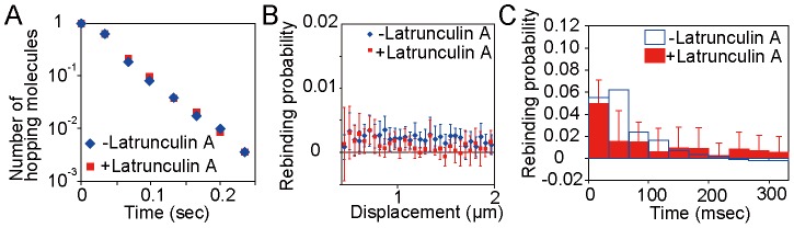
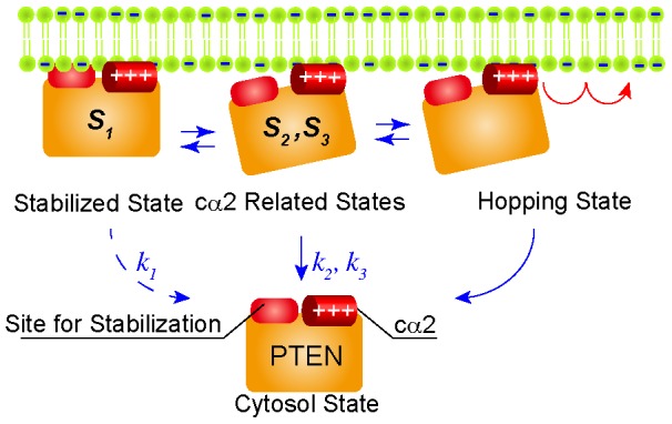
PTEN, a tumor suppressor that is frequently mutated in a wide spectrum of cancers, exerts PI(3,4,5)P3 phosphatase activities that are regulated by its dynamic shuttling between the membrane and cytoplasm. Direct observation of PTEN in the interfacial environment can offer quantitative information about the shuttling dynamics, but remains elusive. Here we show that positively charged residues located in the cα2 helix of the C2 domain are necessary for the membrane localization of PTEN via stable electrostatic interactions in Dictyostelium discoideum. Single-molecule imaging analyses revealed that PTEN molecules moved distances much larger than expected had they been caused by lateral diffusion, a phenomenon we call "hopping." Our novel single-particle tracking analysis method found that the cα2 helix aids in regulating the hopping and stable-binding states. The dynamically established membrane localization of PTEN was revealed to be essential for developmental processes and clarified a fundamental regulation mechanism of the protein quantity and activity on the plasma membrane.
Conflict of interest statement
The authors have declared that no competing interests exist.
Figures


Similar articles
-
Mechanism of human PTEN localization revealed by heterologous expression in Dictyostelium.Oncogene. 2014 Dec 11;33(50):5688-96. doi: 10.1038/onc.2013.507. Epub 2013 Dec 2. Oncogene. 2014. PMID: 24292679 Free PMC article.
-
Defining the membrane-associated state of the PTEN tumor suppressor protein.Biophys J. 2013 Feb 5;104(3):613-21. doi: 10.1016/j.bpj.2012.12.002. Biophys J. 2013. PMID: 23442912 Free PMC article.
-
Engineering PTEN function: membrane association and activity.Methods. 2015 May;77-78:119-24. doi: 10.1016/j.ymeth.2014.10.018. Epub 2014 Oct 22. Methods. 2015. PMID: 25448479 Free PMC article.
-
Membrane association of the PTEN tumor suppressor: neutron scattering and MD simulations reveal the structure of protein-membrane complexes.Methods. 2015 May;77-78:136-46. doi: 10.1016/j.ymeth.2014.10.014. Epub 2014 Oct 27. Methods. 2015. PMID: 25461777 Free PMC article. Review.
-
Structural Mechanisms of PTEN Regulation.Cold Spring Harb Perspect Med. 2020 Mar 2;10(3):a036152. doi: 10.1101/cshperspect.a036152. Cold Spring Harb Perspect Med. 2020. PMID: 31636093 Free PMC article. Review.
Cited by
-
Computational framework for single-cell spatiotemporal dynamics of optogenetic membrane recruitment.Cell Rep Methods. 2022 Jul 6;2(7):100245. doi: 10.1016/j.crmeth.2022.100245. eCollection 2022 Jul 18. Cell Rep Methods. 2022. PMID: 35880018 Free PMC article.
-
Overexpressing TPTE2 (TPIP), a homolog of the human tumor suppressor gene PTEN, rescues the abnormal phenotype of the PTEN-/- mutant.Oncotarget. 2018 Apr 20;9(30):21100-21121. doi: 10.18632/oncotarget.24941. eCollection 2018 Apr 20. Oncotarget. 2018. PMID: 29765523 Free PMC article.
-
Dynamic interactions between a membrane binding protein and lipids induce fluctuating diffusivity.Sci Adv. 2017 Jan 20;3(1):e1601871. doi: 10.1126/sciadv.1601871. eCollection 2017 Jan. Sci Adv. 2017. PMID: 28116358 Free PMC article.
-
Domain-to-domain coupling in voltage-sensing phosphatase.Biophys Physicobiol. 2017 Jun 1;14:85-97. doi: 10.2142/biophysico.14.0_85. eCollection 2017. Biophys Physicobiol. 2017. PMID: 28744425 Free PMC article. Review.
-
Application of single-molecule analysis to singularity phenomenon of cells.Biophys Physicobiol. 2024 May 8;21(Supplemental):e211018. doi: 10.2142/biophysico.bppb-v21.s018. eCollection 2024. Biophys Physicobiol. 2024. PMID: 39175861 Free PMC article.
References
-
- Cevc G (1990) Membrane electrostatics. Biochim Biophys Acta 1031: 311–381. - PubMed
-
- Lemmon MA (2008) Membrane recognition by phospholipid-binding domains. Nat Rev Mol Cell Biol 9: 99–111. - PubMed
-
- Sansal I, Sellers WR (2004) The biology and clinical relevance of the PTEN tumor suppressor pathway. J Clin Oncol 22: 2954–2963. - PubMed
-
- Maehama T, Dixon JE (1998) The tumor suppressor, PTEN/MMAC1, dephosphorylates the lipid second messenger, phosphatidylinositol 3,4,5-trisphosphate. J Biol Chem 273: 13375–13378. - PubMed
MeSH terms
Substances
LinkOut - more resources
Full Text Sources
Other Literature Sources
Research Materials
Miscellaneous
