

Pre-embedding immunogold labeling to optimize protein localization at subcellular compartments and membrane microdomains of leukocytes
- PMID: 25211515
- PMCID: PMC4204927
- DOI: 10.1038/nprot.2014.163
Pre-embedding immunogold labeling to optimize protein localization at subcellular compartments and membrane microdomains of leukocytes
Abstract
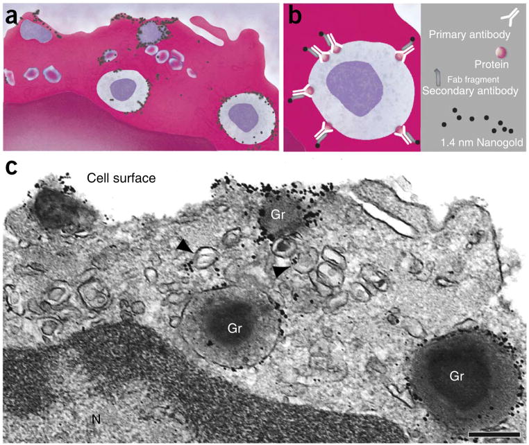
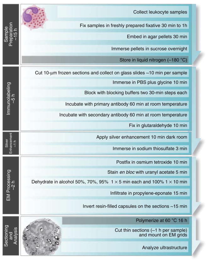
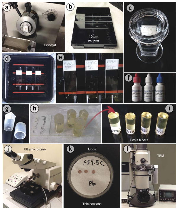
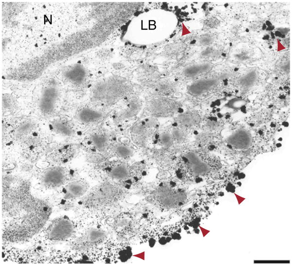
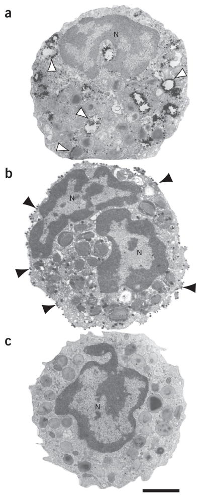
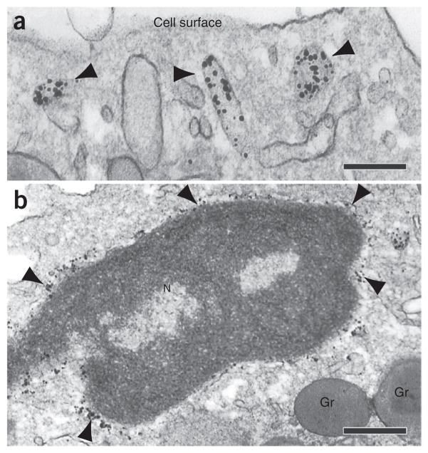
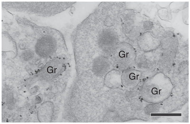
Precise immunolocalization of proteins within a cell is central to understanding cell processes and functions such as intracellular trafficking and secretion of molecules during immune responses. Here we describe a protocol for ultrastructural detection of proteins in leukocytes. The method uses a pre-embedding approach (immunolabeling before standard processing for transmission electron microscopy (TEM)). This protocol combines several strategies for ultrastructure and antigen preservation, robust blocking of nonspecific binding sites, as well as superior antibody penetration for detecting molecules at subcellular compartments and membrane microdomains. A further advantage of this technique is that electron microscopy (EM) processing is quick. This method has been used to study leukocyte biology, and it has helped demonstrate how activated leukocytes deliver specific cargos. It may also potentially be applied to a variety of different cell types. Excluding the initial time required for sample preparation (15 h) and the final resin polymerization step (16 h), the protocol (immunolabeling and EM procedures) can be completed in 8 h.
Conflict of interest statement
Figures
Similar articles
-
Pre-embedding Double-Label Immunoelectron Microscopy of Chemically Fixed Tissue Culture Cells.Methods Mol Biol. 2016;1474:217-32. doi: 10.1007/978-1-4939-6352-2_13. Methods Mol Biol. 2016. PMID: 27515083 Free PMC article.
-
Immunogold labeling of flagellar components in situ.Methods Cell Biol. 2009;91:63-80. doi: 10.1016/S0091-679X(08)91003-7. Epub 2009 Dec 1. Methods Cell Biol. 2009. PMID: 20409780
-
Pre-embedding immunogold localization of antigens in mammalian brain slices.Methods Mol Biol. 2010;657:133-44. doi: 10.1007/978-1-60761-783-9_10. Methods Mol Biol. 2010. PMID: 20602212
-
Pre-embedding labeling for subcellular detection of molecules with electron microscopy.Tissue Cell. 2019 Apr;57:103-110. doi: 10.1016/j.tice.2018.11.002. Epub 2018 Nov 20. Tissue Cell. 2019. PMID: 30497685 Review.
-
Use of ultrastructural immunohistochemistry in human pituitary pathology.Microsc Res Tech. 1992 Jan 15;20(2):152-61. doi: 10.1002/jemt.1070200204. Microsc Res Tech. 1992. PMID: 1547356 Review.
Cited by
-
Trafficking of JC virus-like particles across the blood-brain barrier.Nanoscale Adv. 2021 Feb 9;3(9):2488-2500. doi: 10.1039/d0na00879f. eCollection 2021 May 4. Nanoscale Adv. 2021. PMID: 36134165 Free PMC article.
-
Single-Cell Analyses of Human Eosinophils at High Resolution to Understand Compartmentalization and Vesicular Trafficking of Interferon-Gamma.Front Immunol. 2018 Jul 9;9:1542. doi: 10.3389/fimmu.2018.01542. eCollection 2018. Front Immunol. 2018. PMID: 30038615 Free PMC article.
-
Functions of tissue-resident eosinophils.Nat Rev Immunol. 2017 Dec;17(12):746-760. doi: 10.1038/nri.2017.95. Epub 2017 Sep 11. Nat Rev Immunol. 2017. PMID: 28891557 Free PMC article. Review.
-
Vesicular trafficking of immune mediators in human eosinophils revealed by immunoelectron microscopy.Exp Cell Res. 2016 Oct 1;347(2):385-90. doi: 10.1016/j.yexcr.2016.08.016. Epub 2016 Aug 22. Exp Cell Res. 2016. PMID: 27562864 Free PMC article. Review.
-
Eosinophil ETosis-Mediated Release of Galectin-10 in Eosinophilic Granulomatosis With Polyangiitis.Arthritis Rheumatol. 2021 Sep;73(9):1683-1693. doi: 10.1002/art.41727. Epub 2021 Aug 11. Arthritis Rheumatol. 2021. PMID: 33750029 Free PMC article.
References
-
- Koster AJ, Klumperman J. Electron microscopy in cell biology: integrating structure and function. Nat Rev Mol Cell Biol. 2003;(suppl):SS6–SS10. - PubMed
-
- Margus H, Padari K, Pooga M. Insights into cell entry and intracellular trafficking of peptide and protein drugs provided by electron microscopy. Adv Drug Deliv Rev. 2013;65:1031–1038. - PubMed
-
- Dvorak A, Monahan-Earley R. Diagnostic Ultrastructural Pathology. Vol. 1. CRC Press; 1992.
Publication types
MeSH terms
Substances
Grants and funding
LinkOut - more resources
Full Text Sources
Other Literature Sources
