

Medial prefrontal cortical estradiol rapidly alters memory system bias in female rats: ultrastructural analysis reveals membrane-associated estrogen receptors as potential mediators
- PMID: 25211590
- PMCID: PMC4197985
- DOI: 10.1210/en.2014-1463
Medial prefrontal cortical estradiol rapidly alters memory system bias in female rats: ultrastructural analysis reveals membrane-associated estrogen receptors as potential mediators
Abstract
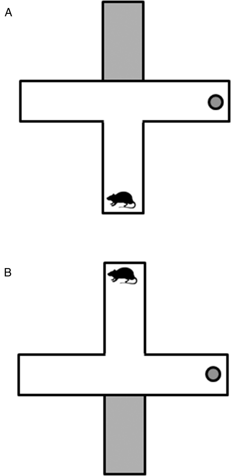
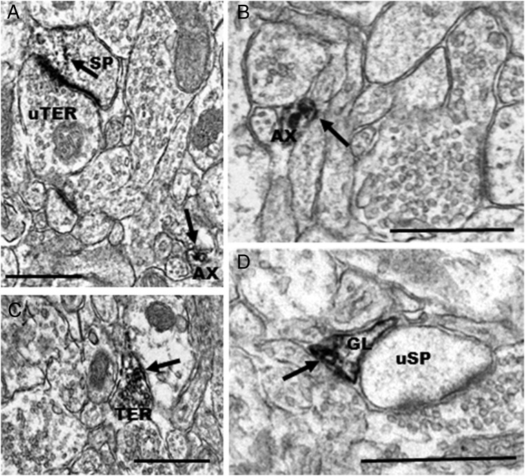
High plasma levels of estradiol (E2) are associated with use of a place memory system over a response memory system. We examined whether infusing estradiol into the medial prefrontal cortex (mPFC) or anterior cingulate cortex (AC) could affect memory system bias in female rats. We also examined the ultrastructural distribution of estrogen receptor (ER)-α, ERβ, and G protein-coupled estrogen receptor 1 (GPER1) in the mPFC of female rats as a mechanism for the behavioral effects of E2 in the mPFC. Each rat was infused bilaterally with either E2 (0.13 μg) or vehicle into the mPFC or AC. The majority of E2 mPFC rats used place memory. In contrast, the majority of mPFC vehicle rats and AC E2 or vehicle rats used response memory. These data show that mPFC E2 rapidly biases females to use place memory. Electron microscopic analysis demonstrated that ERα, ERβ, and GPER1 are localized in the mPFC, almost exclusively at extranuclear sites. This is the first time that GPER1 has been localized to the mPFC of rats and the first time that ERα and ERβ have been described at extranuclear sites in the rat mPFC. The majority of receptors were observed on axons and axon terminals, suggesting that estrogens alter presynaptic transmission in the mPFC. This provides a mechanism via which ERs could rapidly alter transmission in the mPFC to alter PFC-dependent behaviors, such as memory system bias. The discrete nature of immunolabeling for these membrane-associated ERs may explain the discrepancy in previous light microscopy studies.
Figures


References
-
- Tolman EC, Ritchie BF, Kalish D. Studies in spatial learning: orientation and the short-cut. J Exp Psychol. 1946;36:13–24. - PubMed
-
- White NM, McDonald RJ. Multiple parallel memory systems in the brain of the rat. Neurobiol Learn Mem. 2002;77:125–184. - PubMed
-
- Korol DL. Role of estrogen in balancing contributions from multiple memory systems. Neurobiol Learn Mem. 2004;82:309–323. - PubMed
-
- Hussain D, Hoehne A, Woodside B, Brake WG. Reproductive experience modifies the effects of estradiol on learning and memory bias in female rats. Horm Behav. 2013;63:418–423. - PubMed
-
- Korol DL, Kolo LL. Estrogen-induced changes in place and response learning in young adult female rats. Behav Neurosci. 2002;116:411–420. - PubMed
Publication types
MeSH terms
Substances
Grants and funding
LinkOut - more resources
Full Text Sources
Other Literature Sources
Medical
Miscellaneous
