

Protein modification by adenine propenal
- PMID: 25211669
- PMCID: PMC4203390
- DOI: 10.1021/tx500218g
Protein modification by adenine propenal
Abstract
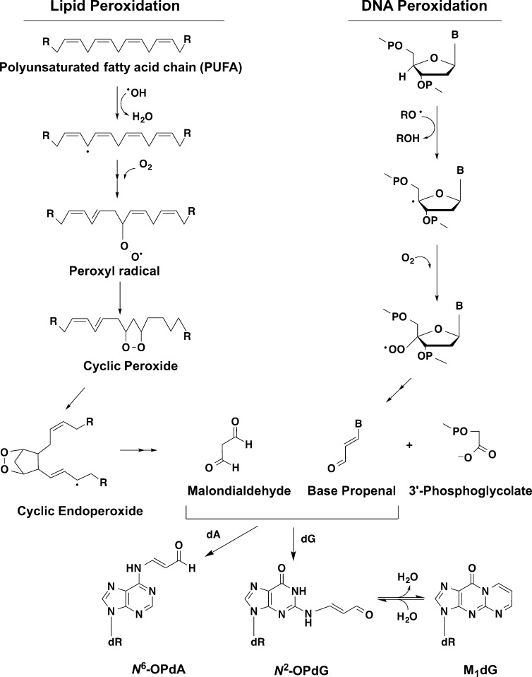
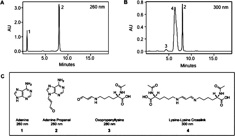
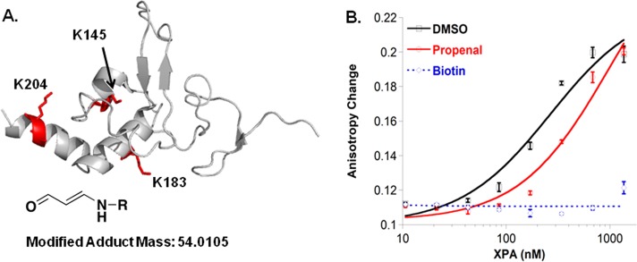
Base propenals are products of the reaction of DNA with oxidants such as peroxynitrite and bleomycin. The most reactive base propenal, adenine propenal, is mutagenic in Escherichia coli and reacts with DNA to form covalent adducts; however, the reaction of adenine propenal with protein has not yet been investigated. A survey of the reaction of adenine propenal with amino acids revealed that lysine and cysteine form adducts, whereas histidine and arginine do not. N(ε)-Oxopropenyllysine, a lysine-lysine cross-link, and S-oxopropenyl cysteine are the major products. Comprehensive profiling of the reaction of adenine propenal with human serum albumin and the DNA repair protein, XPA, revealed that the only stable adduct is N(ε)-oxopropenyllysine. The most reactive sites for modification in human albumin are K190 and K351. Three sites of modification of XPA are in the DNA-binding domain, and two sites are subject to regulatory acetylation. Modification by adenine propenal dramatically reduces XPA's ability to bind to a DNA substrate.
Figures


Similar articles
-
Reaction of malondialdehyde-DNA adducts with hydrazines-development of a facile assay for quantification of malondialdehyde equivalents in DNA.Chem Res Toxicol. 2002 Mar;15(3):312-8. doi: 10.1021/tx010105v. Chem Res Toxicol. 2002. PMID: 11896677
-
Protein modification by lipid peroxidation products: formation of malondialdehyde-derived N(epsilon)-(2-propenol)lysine in proteins.Arch Biochem Biophys. 1997 Oct 1;346(1):45-52. doi: 10.1006/abbi.1997.0266. Arch Biochem Biophys. 1997. PMID: 9328283
-
Reactivity and mutagenicity of endogenous DNA oxopropenylating agents: base propenals, malondialdehyde, and N(epsilon)-oxopropenyllysine.Chem Res Toxicol. 2000 Dec;13(12):1235-42. doi: 10.1021/tx0001631. Chem Res Toxicol. 2000. PMID: 11123964
-
XPA: A key scaffold for human nucleotide excision repair.DNA Repair (Amst). 2016 Aug;44:123-135. doi: 10.1016/j.dnarep.2016.05.018. Epub 2016 May 20. DNA Repair (Amst). 2016. PMID: 27247238 Free PMC article. Review.
-
Three-dimensional structural views of damaged-DNA recognition: T4 endonuclease V, E. coli Vsr protein, and human nucleotide excision repair factor XPA.Mutat Res. 2000 Aug 30;460(3-4):257-75. doi: 10.1016/s0921-8777(00)00031-8. Mutat Res. 2000. PMID: 10946233 Review.
Cited by
-
Intracellular Formation of a DNA Damage-Induced, Histone Post-Translational Modification Following Bleomycin Treatment.J Am Chem Soc. 2022 May 4;144(17):7600-7605. doi: 10.1021/jacs.2c02880. Epub 2022 Apr 25. J Am Chem Soc. 2022. PMID: 35467863 Free PMC article.
-
Histone Adduction and Its Functional Impact on Epigenetics.Chem Res Toxicol. 2017 Jan 17;30(1):376-387. doi: 10.1021/acs.chemrestox.6b00379. Epub 2016 Dec 20. Chem Res Toxicol. 2017. PMID: 27930886 Free PMC article.
-
Covalent Modification of Bromodomain Proteins by Peptides Containing a DNA Damage-Induced, Histone Post-Translational Modification.Chembiochem. 2022 Nov 18;23(22):e202200373. doi: 10.1002/cbic.202200373. Epub 2022 Oct 26. Chembiochem. 2022. PMID: 36173930 Free PMC article.
-
Characterization of covalent modifications of HDL apoproteins by endogenous oxidized phospholipids.Free Radic Biol Med. 2018 Feb 1;115:57-67. doi: 10.1016/j.freeradbiomed.2017.11.012. Epub 2017 Nov 15. Free Radic Biol Med. 2018. PMID: 29155052 Free PMC article.
-
Reactive Carbonyl Species Scavengers-Novel Therapeutic Approaches for Chronic Diseases.Curr Pharmacol Rep. 2017 Apr;3(2):51-67. doi: 10.1007/s40495-017-0081-6. Epub 2017 Feb 14. Curr Pharmacol Rep. 2017. PMID: 28993795 Free PMC article.
References
-
- Hartley D. P.; Kolaja K. L.; Reichard J.; Petersen D. R. (1999) 4-Hydroxynonenal and malondialdehyde hepatic protein adducts in rats treated with carbon tetrachloride: immunochemical detection and lobular localization. Toxicol. Appl. Pharmacol. 161, 23–33. - PubMed
Publication types
MeSH terms
Substances
Grants and funding
- R37 CA087819/CA/NCI NIH HHS/United States
- GM15431/GM/NIGMS NIH HHS/United States
- F32 CA159701-01/CA/NCI NIH HHS/United States
- P01 CA092584/CA/NCI NIH HHS/United States
- T32 ES007028/ES/NIEHS NIH HHS/United States
- P30 DK020593/DK/NIDDK NIH HHS/United States
- S10 RR019022/RR/NCRR NIH HHS/United States
- R01ES1065561/ES/NIEHS NIH HHS/United States
- P30 ES000267/ES/NIEHS NIH HHS/United States
- T32 CA009582/CA/NCI NIH HHS/United States
- DK020593/DK/NIDDK NIH HHS/United States
- P01 GM015431/GM/NIGMS NIH HHS/United States
LinkOut - more resources
Full Text Sources
Other Literature Sources
