

Chronic L-dopa decreases serotonin neurons in a subregion of the dorsal raphe nucleus
- PMID: 25212217
- PMCID: PMC4201267
- DOI: 10.1124/jpet.114.218966
Chronic L-dopa decreases serotonin neurons in a subregion of the dorsal raphe nucleus
Abstract
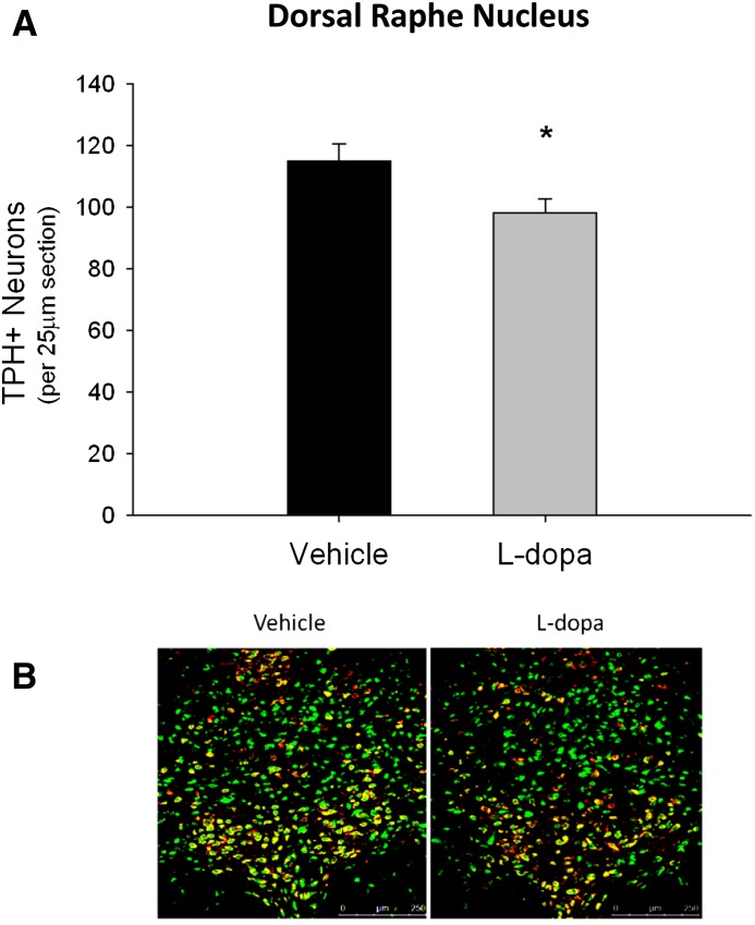
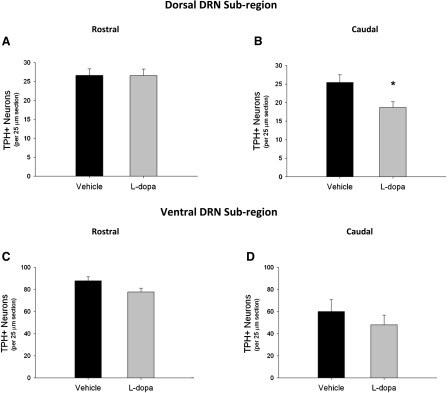
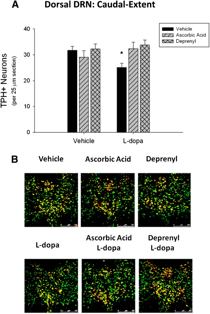
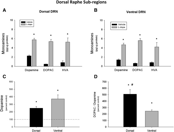
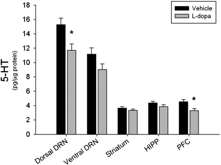
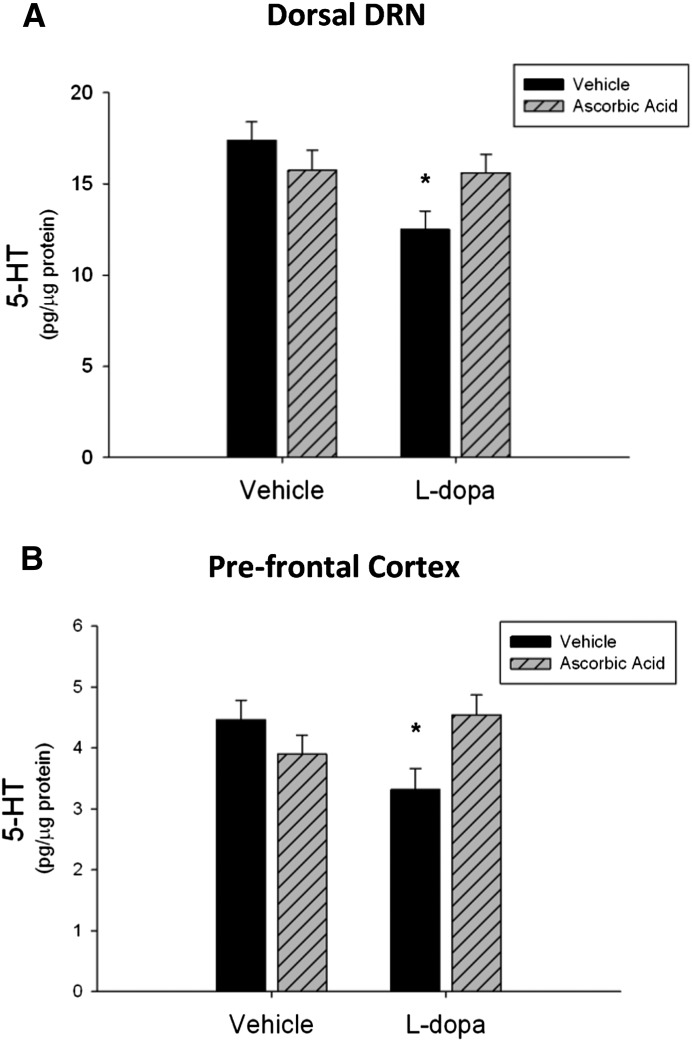
L-Dopa (l-3,4-dihydroxyphenylalanine) is the precursor to dopamine and has become the mainstay therapeutic treatment for Parkinson's disease. Chronic L-dopa is administered to recover motor function in Parkinson's disease patients. However, drug efficacy decreases over time, and debilitating side effects occur, such as dyskinesia and mood disturbances. The therapeutic effect and some of the side effects of L-dopa have been credited to its effect on serotonin (5-HT) neurons. Given these findings, it was hypothesized that chronic L-dopa treatment decreases 5-HT neurons in the dorsal raphe nucleus (DRN) and the content of 5-HT in forebrain regions in a manner that is mediated by oxidative stress. Rats were treated chronically with l-dopa (6 mg/kg; twice daily) for 10 days. Results indicated that the number of 5-HT neurons was significantly decreased in the DRN after l-dopa treatment compared with vehicle. This effect was more pronounced in the caudal-extent of the dorsal DRN, a subregion found to have a significantly higher increase in the 3,4-dihydroxyphenylacetic acid/dopamine ratio in response to acute L-dopa treatment. Furthermore, pretreatment with ascorbic acid (400 mg/kg) or deprenyl (2 mg/kg) prevented the l-dopa-induced decreases in 5-HT neurons. In addition, 5-HT content was decreased significantly in the DRN and prefrontal cortex by l-dopa treatment, effects that were prevented by ascorbic acid pretreatment. Taken together, these data illustrate that chronic L-dopa causes a 5-HT neuron loss and the depletion of 5-HT content in a subregion of the DRN as well as in the frontal cortex through an oxidative-stress mechanism.
Copyright © 2014 by The American Society for Pharmacology and Experimental Therapeutics.
Figures
Similar articles
-
Behavioral impairments and serotonin reductions in rats after chronic L-dopa.Psychopharmacology (Berl). 2015 Sep;232(17):3203-13. doi: 10.1007/s00213-015-3980-4. Epub 2015 Jun 3. Psychopharmacology (Berl). 2015. PMID: 26037945 Free PMC article.
-
The role of the dorsal raphe nucleus in the development, expression, and treatment of L-dopa-induced dyskinesia in hemiparkinsonian rats.Synapse. 2009 Jul;63(7):610-20. doi: 10.1002/syn.20630. Synapse. 2009. PMID: 19309758 Free PMC article.
-
L-DOPA-induced dysregulation of extrastriatal dopamine and serotonin and affective symptoms in a bilateral rat model of Parkinson's disease.Neuroscience. 2012 Aug 30;218:243-56. doi: 10.1016/j.neuroscience.2012.05.052. Epub 2012 Jun 1. Neuroscience. 2012. PMID: 22659568 Free PMC article.
-
Parkinson's disease--opportunities for novel therapeutics to reduce the problems of levodopa therapy.Prog Brain Res. 2008;172:479-94. doi: 10.1016/S0079-6123(08)00923-0. Prog Brain Res. 2008. PMID: 18772047 Review.
-
L-Dopa and Brain Serotonin System Dysfunction.Toxics. 2015 Mar 5;3(1):75-88. doi: 10.3390/toxics3010075. Toxics. 2015. PMID: 29056652 Free PMC article. Review.
Cited by
-
Circuit Mechanisms of L-DOPA-Induced Dyskinesia (LID).Front Neurosci. 2021 Mar 10;15:614412. doi: 10.3389/fnins.2021.614412. eCollection 2021. Front Neurosci. 2021. PMID: 33776634 Free PMC article. Review.
-
Monoaminergic and Histaminergic Strategies and Treatments in Brain Diseases.Front Neurosci. 2016 Nov 24;10:541. doi: 10.3389/fnins.2016.00541. eCollection 2016. Front Neurosci. 2016. PMID: 27932945 Free PMC article. Review.
-
Striatal serotonin transporter gain-of-function in L-DOPA-treated, hemi-parkinsonian rats.Brain Res. 2023 Jul 15;1811:148381. doi: 10.1016/j.brainres.2023.148381. Epub 2023 Apr 29. Brain Res. 2023. PMID: 37127174 Free PMC article.
-
Depression of Serotonin Synaptic Transmission by the Dopamine Precursor L-DOPA.Cell Rep. 2015 Aug 11;12(6):944-54. doi: 10.1016/j.celrep.2015.07.005. Epub 2015 Jul 30. Cell Rep. 2015. PMID: 26235617 Free PMC article.
-
The acute and long-term L-DOPA effects are independent from changes in the activity of dorsal raphe serotonergic neurons in 6-OHDA lesioned rats.Br J Pharmacol. 2016 Jul;173(13):2135-46. doi: 10.1111/bph.13447. Epub 2016 Mar 8. Br J Pharmacol. 2016. PMID: 26805402 Free PMC article.
References
-
- Amat J, Baratta MV, Paul E, Bland ST, Watkins LR, Maier SF. (2005) Medial prefrontal cortex determines how stressor controllability affects behavior and dorsal raphe nucleus. Nat Neurosci 8:365–371 - PubMed
-
- Arai R, Karasawa N, Geffard M, Nagatsu T, Nagatsu I. (1994) Immunohistochemical evidence that central serotonin neurons produce dopamine from exogenous L-DOPA in the rat, with reference to the involvement of aromatic l-amino acid decarboxylase. Brain Res 667:295–299 - PubMed
-
- Bertler A, Falck B, Rosengren E. (1963) The Direct Demonstration of a Barrier Mechanism in the Brain Capillaries. Acta Pharmacol Toxicol (Copenh) 20:317–321 - PubMed
Publication types
MeSH terms
Substances
Grants and funding
LinkOut - more resources
Full Text Sources
Other Literature Sources
