. 2015 Jan;36(1):241-8.
doi: 10.1016/j.neurobiolaging.2014.08.012. Epub 2014 Aug 16.

BAG3 facilitates the clearance of endogenous tau in primary neurons

Affiliations

BAG3 facilitates the clearance of endogenous tau in primary neurons

Zhinian Lei et al. Neurobiol Aging. 2015 Jan.

Abstract

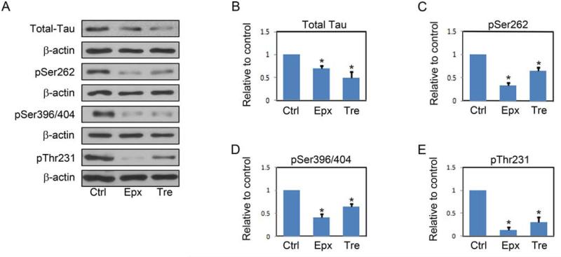
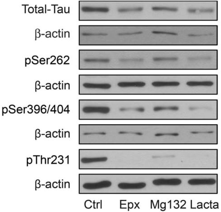
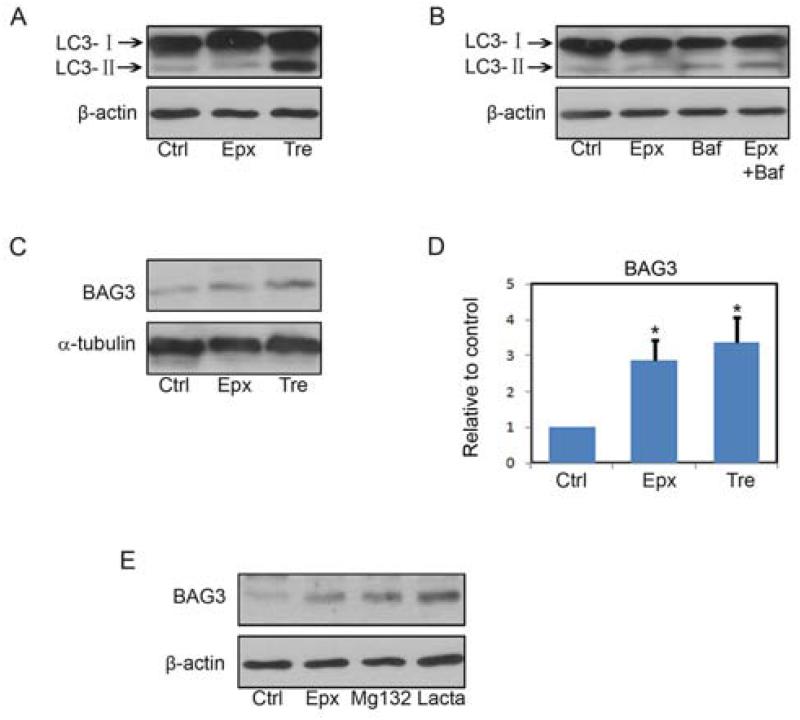
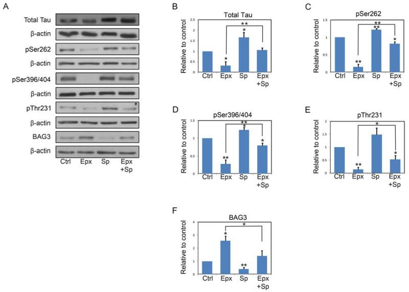
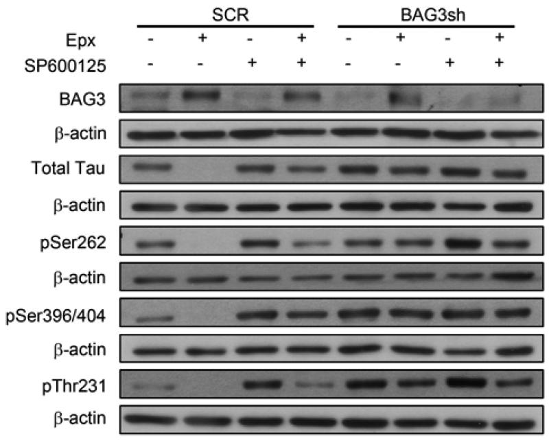
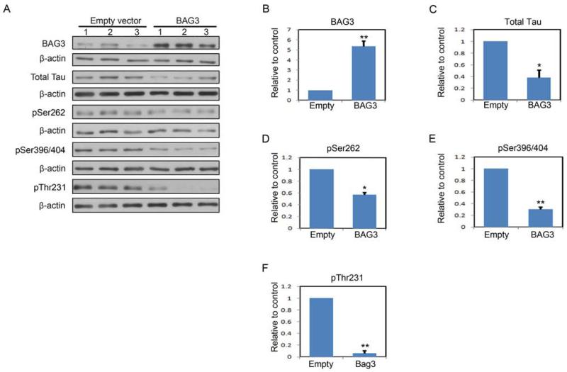
Tau is a microtubule associated protein that is found primarily in neurons, and in pathologic conditions, such as Alzheimer's disease (AD) it accumulates and contributes to the disease process. Because tau plays a fundamental role in the pathogenesis of AD and other tauopathies, and in AD mouse models reducing tau levels improves outcomes, approaches that facilitate tau clearance are being considered as therapeutic strategies. However, fundamental to the development of such interventions is a clearer understanding of the mechanisms that regulate tau clearance. Here, we report a novel mechanism of tau degradation mediated by the co-chaperone BAG3. BAG3 has been shown to be an essential component of a complex that targets substrates to the autophagy pathway for degradation. In rat primary neurons, activation of autophagy by inhibition of proteasome activity or treatment with trehalose resulted in significant decreases in tau and phospho-tau levels. These treatments also induced an upregulation of BAG3. Proteasome inhibition activated JNK, which was responsible for the upregulation of BAG3 and increased tau clearance. Inhibiting JNK or knocking down BAG3 blocked the proteasome inhibition-induced decreases in tau. Further, BAG3 overexpression alone resulted in significant decreases in tau and phospho-tau levels in neurons. These results indicate that BAG3 plays a critical role in regulating the levels of tau in neurons, and interventions that increase BAG3 levels could provide a therapeutic approach in the treatment of AD.

Keywords: Alzheimer's disease; Autophagy; BAG3; Co-chaperone; JNK; Tau.


Figures

FIGURE 1
FIGURE 1. Inhibition of the proteasome by epoxomicin or increasing autophagy with trehalose decreases tau levels
A, Representative immunoblots of lysates from DIV7 primary neurons treated with vehicle only, epoxomicin (Epx, 8nM), or trehalose (Tre, 150mM) for 24 h.. Collected lysates were immunoblotted for total tau and for phosphorylated tau species. For phospho-tau, the 12E8 (pSer262), PHF-1 (pSer396/404) and AT-180 (pThr231) antibodies were used. The membranes were reprobed with a β-actin antibody as a loading control. B-E, shown is quantification of relative levels of tau remaining after treatment described in A. The amount of tau was normalized to β-actin levels and then expressed as the percentage compared with control. Data points are the mean±S.E.M from four independent experiments. *P<0.05.
FIGURE 2
FIGURE 2. Other typical proteasome inhibitors decrease tau levels
Representative immunoblots of lysates from DIV7 primary neurons treated with epoxomicin (Epx, 8nM), MG132 (0.5μM), lactacystin (Lacta, 10μM) or vehicle for 24 h. Collected lysates were immunoblotted for total tau and phosphorylated tau species. For phospho-tau the 12E8 (pSer262), PHF-1 (pSer396/404) and AT-180 (pThr231) antibodies were used. The membranes were also reprobed with a β-actin antibody as a loading control.
FIGURE 3
FIGURE 3. Both epoxomicin and trehalose do not significantly affect neuronal survival
DIV7 Rat primary neurons were treated with epoxomicin (Epx, 8nM) or trehalose (Tre, 150mM) for 24 h and viability was determined with the resazurin (A) or calcein-AM (B) assay as described under “Materials and Methods.” For resazurin assay n=5, for calcein-AM assay n=3.
FIGURE 4
FIGURE 4. Proteasome inhibition and trehalose increase BAG3 levels
A, Rat primary neurons (DIV7) were treated with vehicle, epoxomicin (Epx, 8nM), or trehalose (Tre, 150mM) for 24 h. LC3I/II levels were analyzed by immunoblotting. B, Rat primary neurons (DIV7) were treated with vehicle or epoxomicin (Epx 8nM) for 20 h, Bafilomycin A1 (Baf, 10 nM) was added to the vehicle and epoxomicin treated cells, or no further additions were made and the incubation continued for another 4 h. LC3I/II levels were analyzed by immunoblotting. C, The same treatment as in A but samples were immunoblotted for Bag3. D, Quantitative analysis showed that BAG3 protein levels were significantly increased by both epoxomicin (Epx, 8nM) and trehalose (Tre, 150mM). Bars represent mean±S.E.M. *P<0.05 compared with compared with the control after normalization to the corresponding actin levels. Student’s t-test, n=3. E, Rat primary neurons (DIV7) were treated with vehicle, epoxomicin (Epx 8nM), MG132 (0.5μM) or lactacystin (Lacta, 10μM) for 24 h. BAG3 levels were determined by immunoblotting.
FIGURE 5
FIGURE 5. JNK inhibitor SP600125 decreases the expression of BAG3 and inhibits epoxomicin induced tau clearance
A, Representative immunoblots of lysates from DIV7 primary neurons treated with vehicle only, epoxomicin (Epx, 8nM), SP600125 (Sp, 12.5 μM) or epoxomicin plus SP600125 (Epx+Sp) for 24 h. Collected lysates were immunoblotted for total tau, phosphorylated tau species and BAG3. For phospho-tau, the 12E8 (pSer262), PHF-1 (pSer396/404) and AT-180 (pThr231) antibodies were used. The membranes were reprobed with a β-actin antibody as a loading control. B-F, shown is quantification of relative levels of tau and BAG3 remaining after treatment described in A. The amount of tau was normalized to β-actin levels and then expressed as the percentage compared with control. Data points are the mean±S.E.M from four independent experiments. *P<0.05, **P<0.01.
FIGURE 6
FIGURE 6. BAG3 is required for epoxomicin-induced tau clearance
Representative immunoblots of lysates from DIV7 primary neurons infected with either a scramble (SCR) or BAG3 shRNA virus were treated with vehicle only, epoxomicin (Epx, 8nM), SP600125 (Sp, 12.5 μM) or epoxomicin plus SP600125 (Epx+Sp) for 24 h. Total protein extracts were analyzed for total tau and for phosphorylated tau species. For phospho-tau, the 12E8 (pSer262), PHF-1 (pSer396/404) and AT-180 (pThr231) antibodies were used. Figures are representative of n=3.
FIGURE 7
FIGURE 7. Increased BAG3 results in increased tau clearance
A, Immunoblots of lysates from DIV7 primary neurons infected with either an empty or human BAG3 expression lentivirus vector. Total protein extracts were analyzed for total tau and for phosphorylated tau species. For phospho-tau, the 12E8 (pSer262), PHF-1 (pSer396/404) and AT-180 (pThr231) antibodies were used. B-F, shown is quantification of relative levels of tau remaining after infection described in A. The amount of tau was normalized to β-actin levels and then expressed as the percentage compared with control. Data points are the mean±S.E.M from three independent experiments. *P<0.05, **P<0.01.

Similar articles

Cited by

References

    1. Behl C. BAG3 and friends: co-chaperones in selective autophagy during aging and disease. Autophagy. 2011;7(7):795–8. doi:10.4161/auto.7.7.15844. - PubMed
    1. Boland B, Kumar A, Lee S, Platt FM, Wegiel J, Yu WH, Nixon RA. Autophagy induction and autophagosome clearance in neurons: relationship to autophagic pathology in Alzheimer's disease. The Journal of neuroscience : the official journal of the Society for Neuroscience. 2008;28(27):6926–37. doi:10.1523/JNEUROSCI.0800-08.2008. - PMC - PubMed
    1. Carra S, Seguin SJ, Lambert H, Landry J. HspB8 chaperone activity toward poly(Q)-containing proteins depends on its association with Bag3, a stimulator of macroautophagy. The Journal of biological chemistry. 2008;283(3):1437–44. doi:10.1074/jbc.M706304200. - PubMed
    1. Carrettiero DC, Hernandez I, Neveu P, Papagiannakopoulos T, Kosik KS. The cochaperone BAG2 sweeps paired helical filament-insoluble tau from the microtubule. The Journal of neuroscience : the official journal of the Society for Neuroscience. 2009;29(7):2151–61. doi:10.1523/JNEUROSCI.4660-08.2009. - PMC - PubMed
    1. Chondrogianni N, Gonos ES. Proteasome dysfunction in mammalian aging: steps and factors involved. Experimental gerontology. 2005;40(12):931–8. doi:10.1016/j.exger.2005.09.004. - PubMed

Publication types

MeSH terms