Innervation patterns may limit response to endovascular renal denervation
- PMID: 25212640
- PMCID: PMC4377282
- DOI: 10.1016/j.jacc.2014.07.937
Innervation patterns may limit response to endovascular renal denervation
Abstract
Background: Renal denervation is a new interventional approach to treat hypertension with variable results.
Objectives: The purpose of this study was to correlate response to endovascular radiofrequency ablation of renal arteries with nerve and ganglia distributions. We examined how renal neural network anatomy affected treatment efficacy.
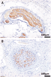
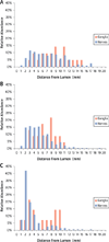
Methods: A multielectrode radiofrequency catheter (15 W/60 s) treated 8 renal arteries (group 1). Arteries and kidneys were harvested 7 days post-treatment. Renal norepinephrine (NEPI) levels were correlated with ablation zone geometries and neural injury. Nerve and ganglion distributions and sizes were quantified at discrete distances from the aorta and were compared with 16 control arteries (group 2).
Results: Nerve and ganglia distributions varied with distance from the aorta (p < 0.001). A total of 75% of nerves fell within a circumferential area of 9.3, 6.3, and 3.4 mm of the lumen and 0.3, 3.0, and 6.0 mm from the aorta. Efficacy (NEPI 37 ng/g) was observed in only 1 of 8 treated arteries where ablation involved all 4 quadrants, reached a depth of 9.1 mm, and affected 50% of nerves. In 7 treated arteries, NEPI levels remained at baseline values (620 to 991 ng/g), ≤20% of the nerves were affected, and the ablation areas were smaller (16.2 ± 10.9 mm(2)) and present in only 1 to 2 quadrants at maximal depths of 3.8 ± 2.7 mm.
Conclusions: Renal denervation procedures that do not account for asymmetries in renal periarterial nerve and ganglia distribution may miss targets and fall below the critical threshold for effect. This phenomenon is most acute in the ostium but holds throughout the renal artery, which requires further definition.
Keywords: aortic tortuosity; renal denervation; superior ostium.
Copyright © 2014 American College of Cardiology Foundation. Published by Elsevier Inc. All rights reserved.
Figures


Comment in
-
Renal denervation for resistant hypertension: not dead yet.J Am Coll Cardiol. 2014 Sep 16;64(11):1088-91. doi: 10.1016/j.jacc.2014.07.947. J Am Coll Cardiol. 2014. PMID: 25212641 No abstract available.
References
-
- Mahfoud F, Ukena C, Schmieder RE, et al. Ambulatory blood pressure changes after renal sympathetic denervation in patients with resistant hypertension. Circulation. 2013;128:132–140. - PubMed
-
- Kaiser L, Beister T, Wiese A, et al. Results of the ALSTER BP real-world registry on renal denervation employing the Symplicity system. Euro- Intervention. 2014;10:157–165. - PubMed
-
- Persu A, Azizi M, Burnier M, Staessen JA. Residual effect of renal denervation in patients with truly resistant hypertension. Hypertension. 2014;62:450–452. - PubMed
-
- Ott C, Mahfoud F, Schmid A, et al. Renal denervation in moderate treatment-resistant hypertension. J Am Coll Cardiol. 2013;62:1880–1886. - PubMed
-
- Esler MD, Krum H, Schlaich M, Schmieder RE, Bohm M, Sobotka PA. Renal sympathetic denervation for treatment of drug-resistant hypertension: one-year results from the Symplicity HTN-2 randomized, controlled trial. Circulation. 2012;126:2976–2982. - PubMed
Publication types
MeSH terms
Grants and funding
LinkOut - more resources
Full Text Sources
Other Literature Sources

