. 2014 Nov;198(3):1071-85.
doi: 10.1534/genetics.114.168294. Epub 2014 Sep 10.

Genetic analysis of the ribosome biogenesis factor Ltv1 of Saccharomyces cerevisiae

Affiliations

Genetic analysis of the ribosome biogenesis factor Ltv1 of Saccharomyces cerevisiae

Jason R Merwin et al. Genetics. 2014 Nov.

Abstract

Ribosome biogenesis has been studied extensively in the yeast Saccharomyces cerevisiae. Yeast Ltv1 is a conserved 40S-associated biogenesis factor that has been proposed to function in small subunit nuclear export. Here we show that Ltv1 has a canonical leucine-rich nuclear export signal (NES) at its extreme C terminus that is both necessary for Crm1 interaction and Ltv1 export. The C terminus of Ltv1 can substitute for the NES in the 60S-export adapter Nmd3, demonstrating that it is a functional NES. Overexpression of an Ltv1 lacking its NES (Ltv1∆C13) was strongly dominant negative and resulted in the nuclear accumulation of RpS3-GFP; however, export of the pre-40S was not affected. In addition, expression of endogenous levels of Ltv1∆C protein complemented both the slow-growth phenotype and the 40S biogenesis defect of an ltv1 deletion mutant. Thus, if Ltv1 is a nuclear export adapter for the pre-40S subunit, its function must be fully redundant with additional export factors. The dominant negative phenotype of Ltv1∆NES overexpression was suppressed by co-overexpressing RpS3 and its chaperone, Yar1, or by deletion of the RpS3-binding site in Ltv1∆NES, suggesting that titration of RpS3 by Ltv1∆NES is deleterious in yeast. The dominant-negative phenotype did not correlate with a decrease in 40S levels but rather with a reduction in the polysome-to-monosome ratio, indicating reduced rates of translation. We suggest that titration of RpS3 by excess nuclear Ltv1 interferes with 40S function or with a nonribosomal function of RpS3.

Keywords: Ltv1; RpS3; nuclear export; ribosome biogenesis; small subunit.


Figures

Figure 1
Figure 1
The C terminus of Ltv1 is necessary for nuclear export. (A) Schematic of Ltv1 C-terminal deletions. Putative NES sequence is indicated, with the consensus hydrophobic residues shaded; potential NLS sequences (PKGRRAH and KKKRK) are also shown. (B) Cell dilution assay. Wild-type (LY134) and ∆ltv1 (LY136) cells were transformed with the following plasmids: empty vector (pUG23) and Ltv1 with GFP fused to the N terminus (Ld144) or the C terminus (Ld47). Stationary-phase cultures were adjusted to an OD660 of 1.0, diluted serially 1:5, spotted onto selective plates, and grown at 30° for 72 hr prior to imaging. (C) Fluorescence microscopy of ∆ltv1 cells transformed with Ltv1-GFP (Ld144), GFP-Ltv1∆C69 (Ld143), and Ltv1∆C237-GFP (Ld119).
Figure 2
Figure 2
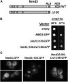
The C terminus of Ltv1 is sufficient for nuclear export. (A) Schematic diagram of Nmd3 indicating the NES and NLS sequences. Nmd3∆100 removes 100 residues including the NES from the C terminus. (B) nmd3-4ts (LY271) cells were transformed with the following plasmids: empty vector (pRS315), wild-type NMD3-GFP (pAJ582), nmd3∆100-GFP (pAJ584), or nmd3∆100-ltv1C71-GFP (nmd3∆100 plus sequence encoding the last 71 amino acids of Ltv1) (Ld165). Equal numbers of cells from each strain were spotted onto selective plates and grown at the indicated temperatures for 60 hr. (C) Wild-type (LY134) cells were transformed with NMD3-GFP (pAJ582), nmd3∆100-GFP (pAJ584), or nmd3∆100-ltv1C71-GFP (Ld165). Live, log-phase cells were imaged by fluorescence microscopy.
Figure 3
Figure 3
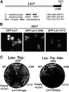
A consensus NES in the C terminus of Ltv1 is necessary for export and Crm1 interaction. (A) Schematic diagram of Ltv1 showing the consensus NES within the region deleted in Ltv1∆C69. The amino acid sequence of the Ltv1 NES is shown aligned with the Nmd3 NES and with the revised NES consensus (Guttler et al. 2010). The consensus has five hydrophobic positions shown as shaded (Ø0–Ø4) and interspersed with a specific number of hydrophilic residues (X), where the subscript refers to the number of residues. (B) Fluorescence microscopy of live, log-phase Δltv1 cells transformed with plasmids expressing N-terminal GFP-tagged Ltv1; GFP-Ltv1 (Ld144), GFP-Ltv1∆C69 (Ld143), and GFP-Ltv1∆C13 (Ld153). Cells were grown into log phase (OD660 0.5–0.8) in selective media at 30° prior to imaging. (C) Two-hybrid assay. Full-length and truncated forms of Ltv1 fused to an N-terminal GAL4 activation domain (Empty, pGADT7g; WT, Ld170; ∆N55, Ld172; ∆C69, Ld174; ∆C13, Ld173) were tested for interaction with Crm1 fused to an N-terminal GAL4 DNA-binding domain (pGBD-Crm1) or empty vector (pGBDC2) (James et al. 1996). The fusion constructs were cotransformed into PJ69a cells containing integrated GAL1::HIS3 and GAL2::ADE2 reporter genes (James et al. 1996). Activation of the ADE2 reporter gene was measured by comparing growth on selective plates lacking leucine and tryptophan (Leu− Trp−) media ± adenine (Ade), as indicated.
Figure 4
Figure 4
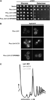
Ltv1∆NES overexpression is dominant negative. (A) Cell dilution assay. Galactose-inducible plasmids expressing Ltv1 (Ld180), Ltv1∆C13 (Ld181), Ltv1∆C69 (Ld182), and Ltv1∆C13PKINES (Ld329) were transformed into wild-type yeast (LY134) as indicated. Transformants were grown overnight in selective media containing glucose. Cells were then adjusted to an OD660 of 1.0, serially diluted 1:5, spotted onto selective plates containing glucose or galactose, and imaged after 48 hr at 30°. (B) RpS3-GFP microscopy. Transformants described in (A) were cotransformed with PRpS3-RpS3-GFP (Ld70) grown in selective media with 2% glucose overnight at 30°, spun down, and resuspended in selective media with 2% raffinose, grown overnight, and finally diluted into selective media with 2% galactose. Log-phase cells were imaged 12 hr later. (C) Polysome profiles of strains described in A. Cultures were incubated overnight in selective media containing glucose, spun down, washed once in selective media with galactose, resuspended to an OD of 0.1 in selective media containing 2% galactose, and incubated for 5 hr until cells reached mid-log phase. Extracts were analyzed on sucrose density gradients as described in Materials and Methods. Traces showing the absorbance of rRNA at 254 nm of each fraction are overlaid.
Figure 5
Figure 5
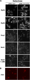
Ltv1∆NES overexpression affects Rps3-GFP, but not pre-40S export. (A) Fluorescence microscopy of cells expressing various GFP-tagged proteins and overexpressing either Ltv1 (pGAL-Ltv1) or Ltv1∆NES (pGAL-Ltv1∆C13) on media containing galactose. For RpS3 and RpS2, wild-type (LY134) cells were cotransformed with Ld140 (PRpS3- RPS3-GFP) or Ld70 (PRpS2-RPS2-GFP) and either PGAL-LTV1 (Ld180) or PGAL-ltv1∆C13 (Ld181). For Enp1, LY256 cells, which have GFP integrated at the ENP1 locus, were transformed with Ld177 (PGAL-LTV1) or Ld178 (PGAL-ltv1∆C13). For Nob1, an LMB-sensitive strain with an integrated NOB1-GFP gene (LY261) was transformed with Ld177 (PGAL-LTV1) or Ld178 (PGAL-ltv1∆C13). All cultures were grown to log phase in selective media with raffinose before dilution into selective media with either 2% galactose or 2% glucose. Cells were grown into log phase (6 hr) before imaging. Cells treated with LMB were incubated for 2 hr with 100 ng/ml LMB at room temperature following growth in 2% galactose. (B) FISH. Wild-type cells (LY134) were transformed with plasmids Ld180 and Ld181. Cultures were grown to stationary phase in selective media containing 2% glucose, diluted into fresh media containing 2% galactose, and grown overnight to an OD660 of 0.4–0.6. A Cy3-labeled DNA probe complementary to the 5′-ITS1 sequence was hybridized to fixed, permeabilized cells prior to imaging using TRITC filters.
Figure 6
Figure 6
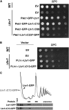
ltv1∆C13 complements the slow-growth and ribosome biogenesis phenotypes of ∆ltv1. (A) Cell dilution assay. Wild-type (LY134) and ∆ltv1 (LY136) cells were transformed with empty vector (pUG34) and ∆ltv1 cells with the following MET25 promoter constructs: GFP-Ltv1 (Ld144), GFP-Ltv1∆C13 (Ld153), GFP-Ltv1∆C69 (Ld143), and Ltv1∆C237-GFP (Ld119). Transformants were grown in selective media overnight, diluted to an OD of 1.0, serially diluted 1:5, and spotted onto selective plates containing 200 µM methionine. Cells were grown at 22° and imaged after 48 hr. (B) Cell dilution assay. Wild-type (LY134) and ∆ltv1 (LY136) cells were transformed with empty vector (pUG35), and ∆ltv1 cells were transformed with Ltv1-GFP (Ld68) or Ltv1∆C13-GFP (Ld183) under the control of the LTV1 promoter. Cells were diluted to an OD600 of 1.0, serially diluted 1:10, and spotted onto selective plates in duplicate. Plates were incubated at 23° for 2 days prior to imaging. (C) Polysome profiles. ∆ltv1 (LY136) cells were transformed with plasmids expressing Ltv1-GFP (Ld68) or Ltv1∆C13-GFP (Ld183) from the LTV1 promoter. Extracts were run on 7–47% sucrose gradients, and fractions were analyzed by SDS-PAGE as described in Materials and Methods. Ltv1 was detected using anti-GFP.
Figure 7
Figure 7
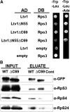
Ltv1∆C and RpS3 interact and coprecipitate. (A) Two-hybrid assay. Vectors encoding full-length and truncated forms of Ltv1 fused with a C-terminal GAL4 activation domain; Ltv1 (Ld235), Ltv1ΔN55 (Ld236), Ltv1ΔC69 (Ld237), Ltv1ΔN55ΔC69 (Ld238), and empty vector (pGADCg) were cotransformed into PJ69a cells with a vector encoding RpS3 fused to an N-terminal GAL4 DNA-binding domain, pGBKT7-Rps3 (Ld233). Transformants were grown overnight to stationary phase, adjusted to an OD660 of 1.0, serially diluted 1:5, and spotted onto selective plates lacking tryptophan and leucine (−Trp, −Leu) or tryptophan, leucine, and adenine (−Trp, −Leu, −Ade), as indicated. (B) Immunoprecipitation. Gal-inducible plasmids expressing wild-type Ltv1-CFP (Ld206) and Ltv1ΔC69-CFP (Ld205) were transformed into wild-type (LY134) cells. Cultures were grown in selective media containing 2% galactose to an OD660 of 0.4–0.6, collected by centrifugation, and lysed. Ltv1-CFP was immunoprecipitated with Epoxy M-270 Dyna-beads coupled to anti-GFP antibody, and Western blot analysis of proteins eluted from the beads was performed with the indicated antibodies. A minus CFP antibody control is indicated by the “Cont” designation.
Figure 8
Figure 8
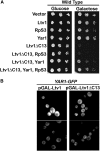
RpS3/Yar1 overexpression rescues the dominant-negative phenotype of Ltv1∆NES over-expression, but Ltv1∆NES does not retain Yar1. (A) Serial dilution assay. Galactose-inducible plasmids expressing full-length Ltv1 (Ld180), Ltv1∆C13 (Ld181), RpS3 (Ld208), and Yar1 (Ld8) were transformed into wild-type yeast (LY134). Cell dilution assays were preformed as described in Figure 4. (B) Yar1-GFP localization. Cells with an integrated Yar1-GFP gene (LY278) were transformed with PGAL-LTV1 (Ld177) or PGAL-LTV1∆C13 (Ld178). Cultures were grown as described in Figure 5 and imaged after 6 hr growth in selective media with 2% galactose.
Figure 9
Figure 9
Deletion of Rps3 binding site in Ltv1∆NES eliminates nuclear retention of Rps3 and rescues its dominant-negative phenotype. (A) Schematic diagram showing extent of internal deletions in LTV1∆C13. The amino acids deleted are identified by their position from the N terminus of Ltv1. The constructs are numbered 1–6, and those that encode proteins that interact with RpS3 are denoted with a “+” in the right column. (B) Two-hybrid assay. PJ69a cells were cotransformed with pGBKT7g-RpS3 (Ld233) and various Ltv1 deletions fused to a C-terminal GAL4 activation domain (AD) as follows: (1) Ltv1∆C13 (Ld263), (2) Ltv1∆56-102∆C13 (Ld269), (3) Ltv1∆103-201∆C13 (Ld270), (4) Ltv1∆202-321∆C13 (Ld259), (5) Ltv1∆322-332∆C13 (Ld261), and (6) Ltv1∆333-394∆C13 (Ld260). C1 is PJ69a cells cotransformed with Ltv1-AD and pGBKT7g, and C2 has pGADCg and pGBKT7g-RpS3 (Ld233). Single colonies were streaked onto selective plates with or without added histidine (−leu −trp and −leu −trp −his, respectively). (C) Fluorescence microscopy of wild-type cells (LY134) transformed with vectors expressing Ltv1-GFP (Ld258), Ltv1∆C13-GFP (Ld257), and Ltv1∆56-102∆C13-GFP (Ld255). Cells were grown in selective media with glucose into log phase before imaging. (D) Fluorescence microscopy of live, log-phase cells (LY134) cotransformed with Ld140 (PRpS3-RpS3-GFP) and galactose-inducible plasmids expressing full-length Ltv1 (Ld180), Ltv1∆C13 (Ld181), or ltv1Δ56-102ΔC13 (Ld276). Cells were induced on galactose as described in Figure 6A prior to imaging. (E) Cultures of strain BY4741 expressing Ltv1-GFP (Ld258), Ltv1∆C13 (Ld257), Ltv1∆56-102∆C13 (Ld255), Gal4AD-Ltv1 (Ld235), Gal4-Ltv1∆C13 (Ld263), or Gal4AD-Ltv1∆C56-102∆C13 (Ld269) were grown to an OD600 of 0.3. Whole-cell extracts were prepared, and 0.3 OD600 units were loaded per lane and separated by SDS-PAGE. Proteins were transferred to membrane and probed for GFP, Gal4 activation domain, or Rpl8 as a loading control. (F) Galactose-inducible plasmids expressing full-length Ltv1 (Ld180), Ltv1∆C13 (Ld181), Ltv1∆56-102 (Ld275), Ltv1Δ56-102ΔC13 (Ld276), or empty vector (pAG413GAL) were transformed into wild-type yeast (LY134) as indicated. Cell dilution assays were preformed as described in Figure 4A.

Comment in

Similar articles

Cited by

References

    1. Alberti S., Gitler A. D., Lindquist S., 2007. A suite of Gateway cloning vectors for high-throughput genetic analysis in Saccharomyces cerevisiae. Yeast 24: 913–919. - PMC - PubMed
    1. Amberg D. C., Goldstein A. L., Cole C. N., 1992. Isolation and characterization of RAT1: an essential gene of Saccharomyces cerevisiae required for the efficient nucleocytoplasmic trafficking of mRNA. Genes Dev. 6: 1173–1189. - PubMed
    1. Bassler J., Grandi P., Gadal O., Lessmann T., Petfalski E., et al. , 2001. Identification of a 60S preribosomal particle that is closely linked to nuclear export. Mol. Cell 8: 517–529. - PubMed
    1. Bradatsch B., Katahira J., Kowalinski E., Bange G., Yao W., et al. , 2007. Arx1 functions as an unorthodox nuclear export receptor for the 60S preribosomal subunit. Mol. Cell 27: 767–779. - PubMed
    1. Campbell M. G., Karbstein K., 2011. Protein-protein interactions within late pre-40S ribosomes. PLoS ONE 6: e16194. - PMC - PubMed

Publication types

MeSH terms