

Coating solid dispersions on microneedles via a molten dip-coating method: development and in vitro evaluation for transdermal delivery of a water-insoluble drug
- PMID: 25213295
- PMCID: PMC4374630
- DOI: 10.1002/jps.24159
Coating solid dispersions on microneedles via a molten dip-coating method: development and in vitro evaluation for transdermal delivery of a water-insoluble drug
Abstract
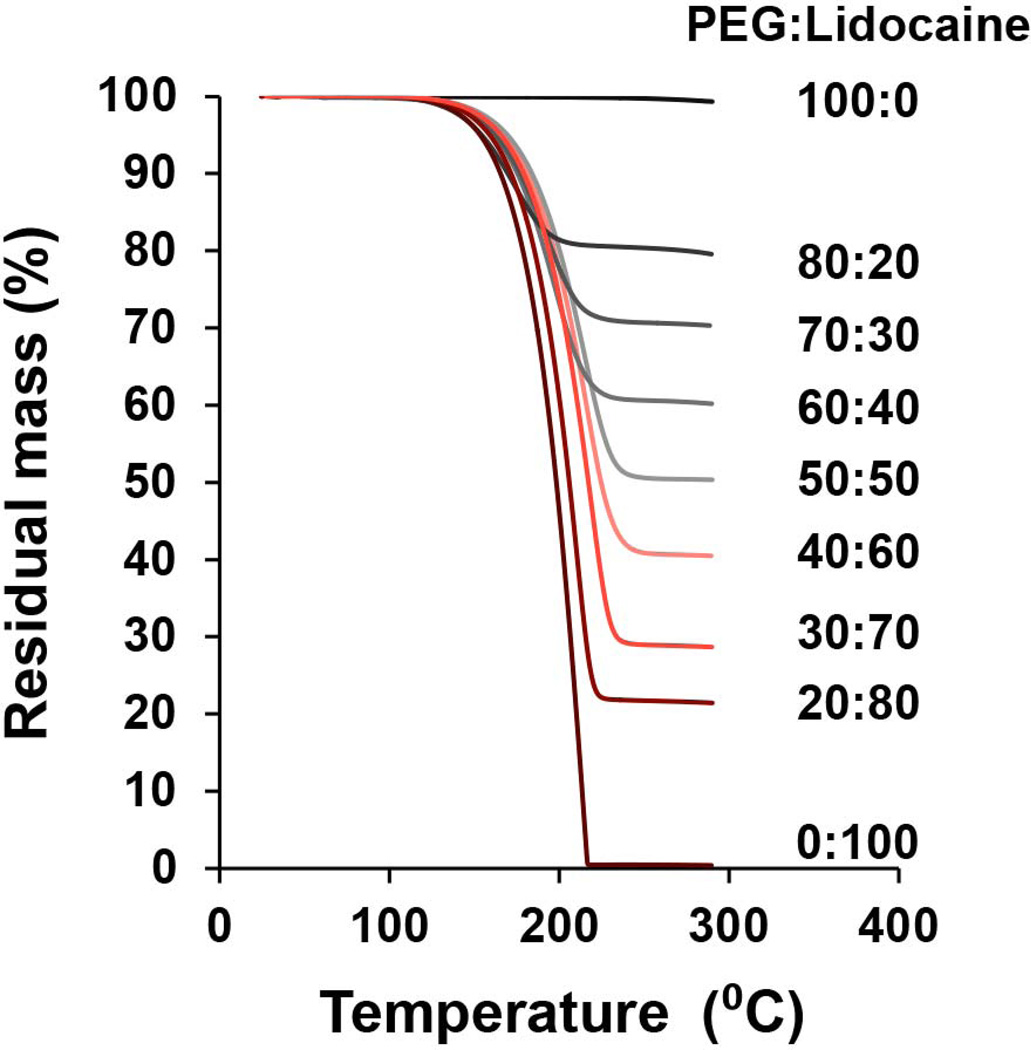
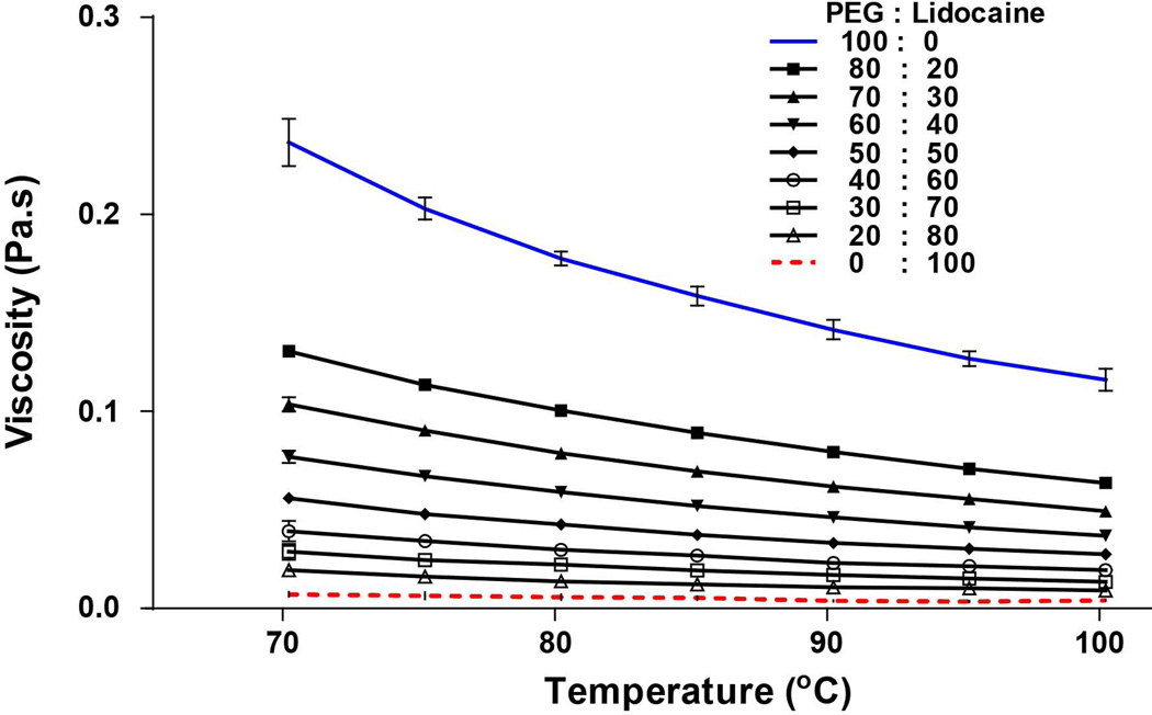
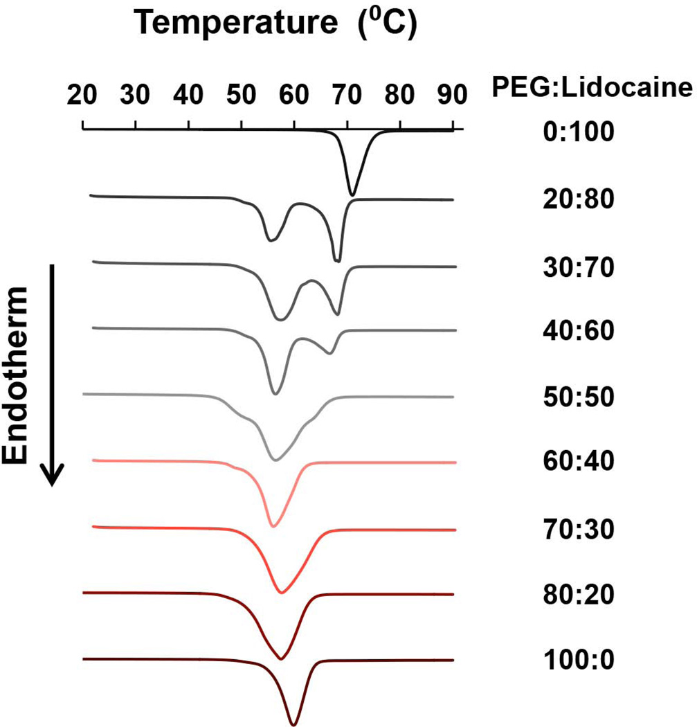
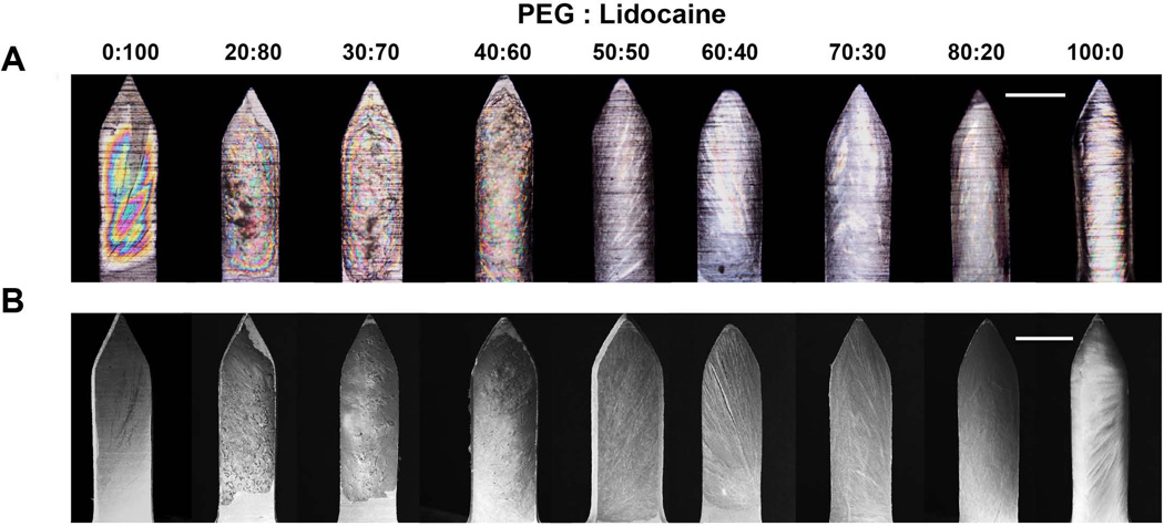
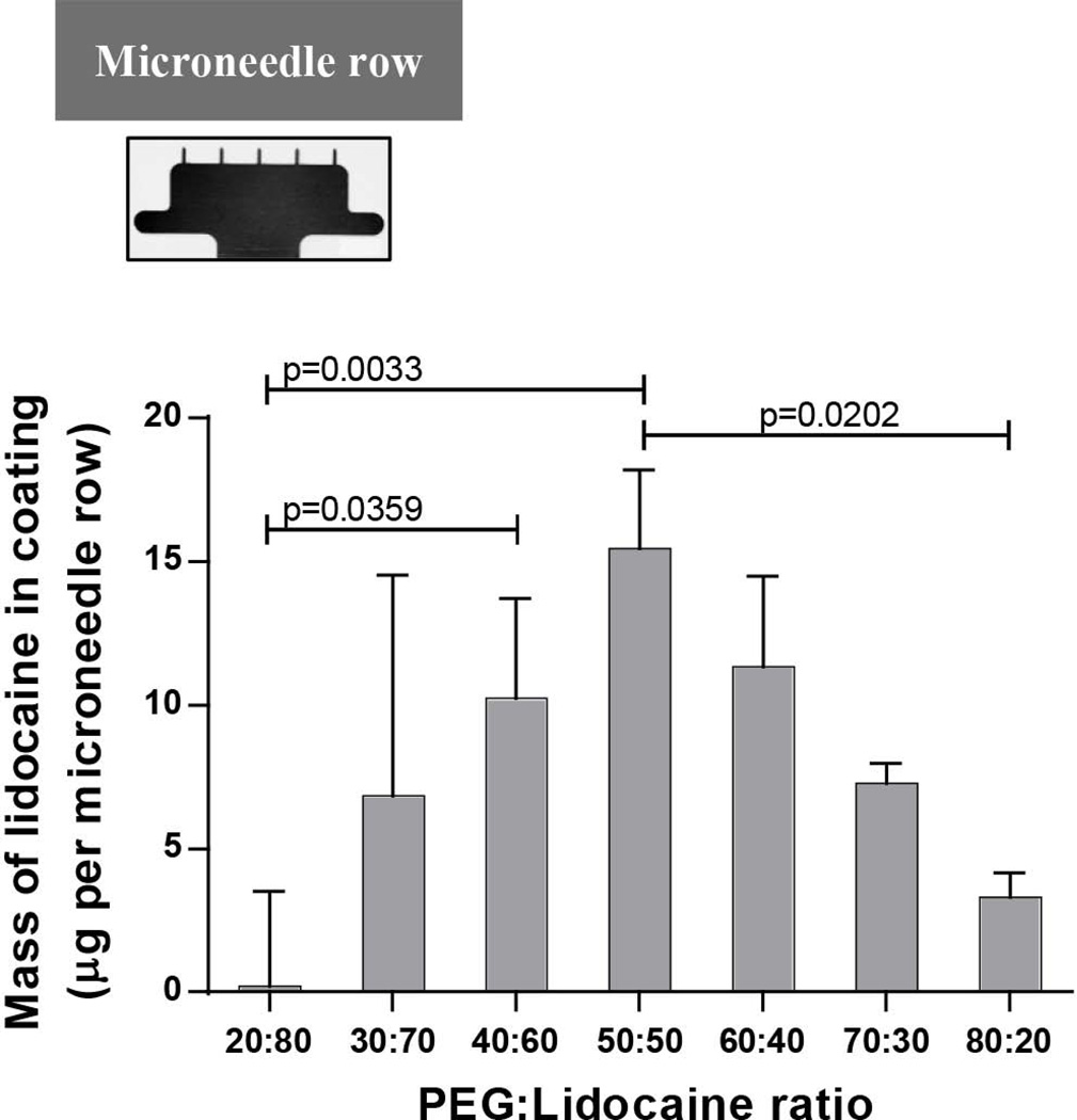
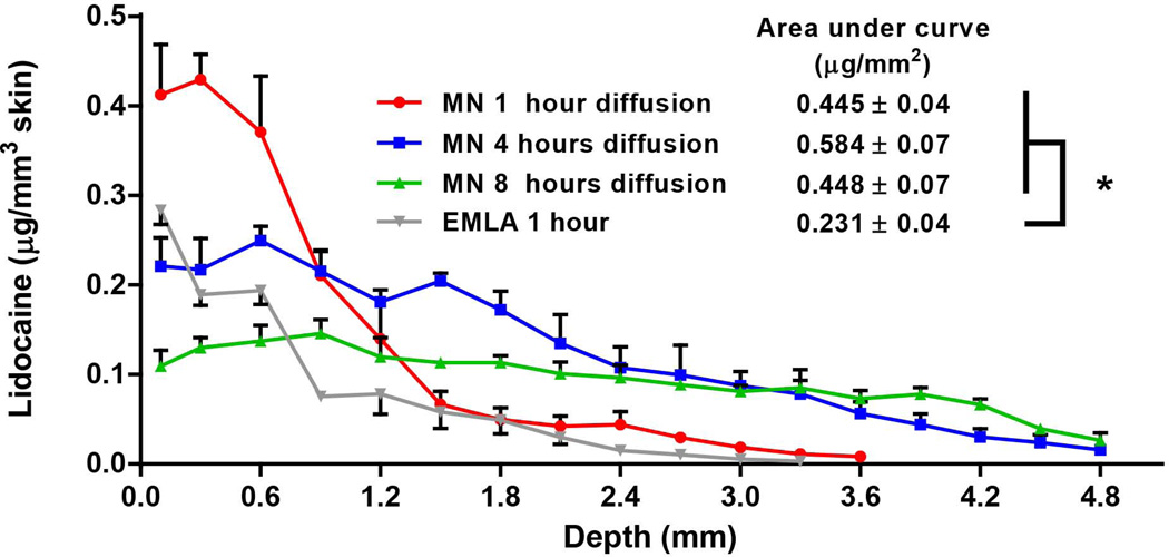
This study demonstrates for the first time the ability to coat solid dispersions on microneedles as a means to deliver water-insoluble drugs through the skin. Polyethylene glycol (PEG) was selected as the hydrophilic matrix, and lidocaine base was selected as the model hydrophobic drug to create the solid dispersion. First, thermal characterization and viscosity measurements of the PEG-lidocaine mixture at different mass fractions were performed. The results show that lidocaine can remain stable at temperatures up to ∼130°C and that viscosity of the PEG-lidocaine molten solution increases as the mass fraction of lidocaine decreases. Differential scanning calorimetry demonstrated that at lidocaine mass fraction less than or equal to 50%, lidocaine is well dispersed in the PEG-lidocaine mixture. Uniform coatings were obtained on microneedle surfaces. In vitro dissolution studies in porcine skin showed that microneedles coated with PEG-lidocaine dispersions resulted in significantly higher delivery of lidocaine in just 3 min compared with 1 h topical application of 0.15 g EMLA®, a commercial lidocaine-prilocaine cream. In conclusion, the molten coating process we introduce here offers a practical approach to coat water-insoluble drugs on microneedles for transdermal delivery.
Keywords: coating; dissolution; solid dispersion; solubility; transdermal drug delivery.
© 2014 Wiley Periodicals, Inc. and the American Pharmacists Association.
Conflict of interest statement
This potential conflict of interest has been disclosed and is managed by Texas Tech University.
Figures

Similar articles
-
Development of lidocaine-coated microneedle product for rapid, safe, and prolonged local analgesic action.Pharm Res. 2012 Jan;29(1):170-7. doi: 10.1007/s11095-011-0524-4. Epub 2011 Jul 7. Pharm Res. 2012. PMID: 21735335
-
Hard polymeric porous microneedles on stretchable substrate for transdermal drug delivery.Sci Rep. 2022 Feb 3;12(1):1853. doi: 10.1038/s41598-022-05912-6. Sci Rep. 2022. PMID: 35115643 Free PMC article.
-
Drug-coated microneedles for rapid and painless local anesthesia.Biomed Microdevices. 2017 Mar;19(1):2. doi: 10.1007/s10544-016-0144-1. Biomed Microdevices. 2017. PMID: 28070698
-
Microneedle Coating Methods: A Review with a Perspective.J Pharmacol Exp Ther. 2019 Sep;370(3):555-569. doi: 10.1124/jpet.119.258707. Epub 2019 Jun 7. J Pharmacol Exp Ther. 2019. PMID: 31175217 Free PMC article. Review.
-
Microneedles for transdermal drug delivery.Adv Drug Deliv Rev. 2004 Mar 27;56(5):581-7. doi: 10.1016/j.addr.2003.10.023. Adv Drug Deliv Rev. 2004. PMID: 15019747 Review.
Cited by
-
Microneedle-Mediated Transdermal Delivery of Biopharmaceuticals.Pharmaceutics. 2023 Jan 13;15(1):277. doi: 10.3390/pharmaceutics15010277. Pharmaceutics. 2023. PMID: 36678906 Free PMC article. Review.
-
Design and Development of Liquid Drug Reservoirs for Microneedle Delivery of Poorly Soluble Drug Molecules.Pharmaceutics. 2019 Nov 13;11(11):605. doi: 10.3390/pharmaceutics11110605. Pharmaceutics. 2019. PMID: 31766145 Free PMC article.
-
Recent advances of controlled drug delivery using microfluidic platforms.Adv Drug Deliv Rev. 2018 Mar 15;128:3-28. doi: 10.1016/j.addr.2017.09.013. Epub 2017 Sep 15. Adv Drug Deliv Rev. 2018. PMID: 28919029 Free PMC article. Review.
-
The Role of Natural Deep Eutectic Solvents in a Hydrogel Formulation Containing Lidocaine.Pharmaceutics. 2025 Mar 2;17(3):324. doi: 10.3390/pharmaceutics17030324. Pharmaceutics. 2025. PMID: 40142988 Free PMC article.
-
A Semi-Dissolving Microneedle Patch Incorporating TEMPO-Oxidized Bacterial Cellulose Nanofibers for Enhanced Transdermal Delivery.Polymers (Basel). 2020 Aug 20;12(9):1873. doi: 10.3390/polym12091873. Polymers (Basel). 2020. PMID: 32825232 Free PMC article.
References
-
- van der Maaden K, Jiskoot W, Bouwstra J. Microneedle technologies for (trans)dermal drug and vaccine delivery. J Control Release. 2012;161(2):645–655. - PubMed
-
- Zhang Y, Brown K, Siebenaler K, Determan A, Dohmeier D, Hansen K. Development of Lidocaine-Coated Microneedle Product for Rapid, Safe, and Prolonged Local Analgesic Action. Pharm Res. 2012;29(1):170–177. - PubMed
Publication types
MeSH terms
Substances
Grants and funding
LinkOut - more resources
Full Text Sources
Other Literature Sources
