. 2015 Mar 1;105(3):248-59.
doi: 10.1093/cvr/cvu207. Epub 2014 Sep 11.

Emergence of Orai3 activity during cardiac hypertrophy

Affiliations

Emergence of Orai3 activity during cardiac hypertrophy

Youakim Saliba et al. Cardiovasc Res. .

Abstract

Aims: Stromal interaction molecule 1 (STIM1) has been shown to control a calcium (Ca(2+)) influx pathway that emerges during the hypertrophic remodelling of cardiomyocytes. Our aim was to determine the interaction of Orai1 and Orai3 with STIM1 and their role in the constitutive store-independent and the store-operated, STIM1-dependent, Ca(2+) influx in cardiomyocytes.

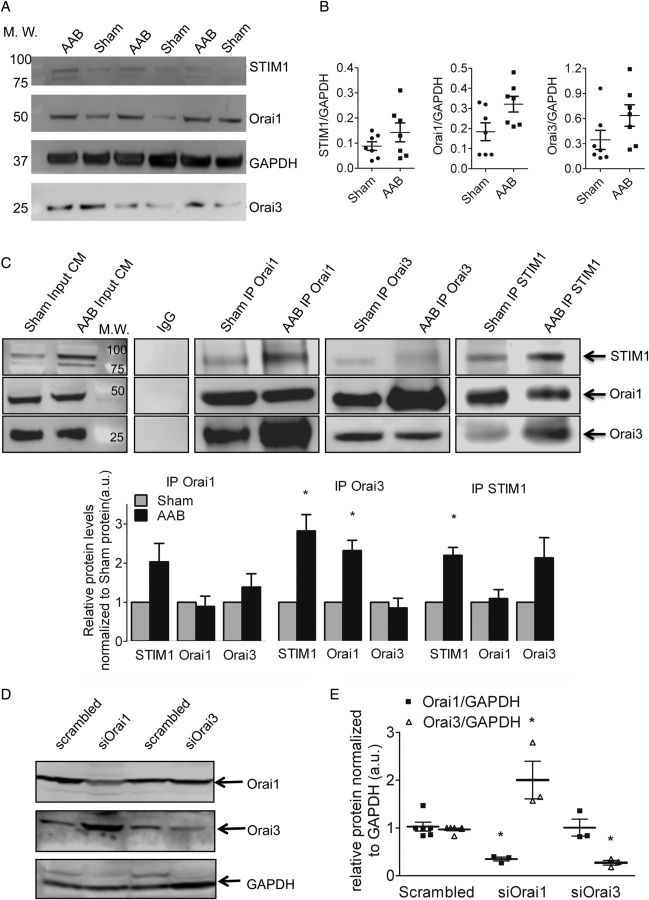
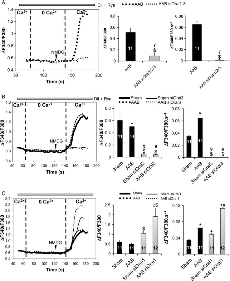
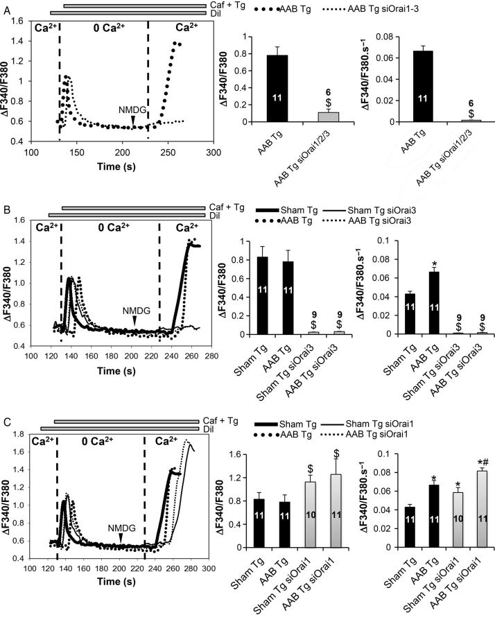
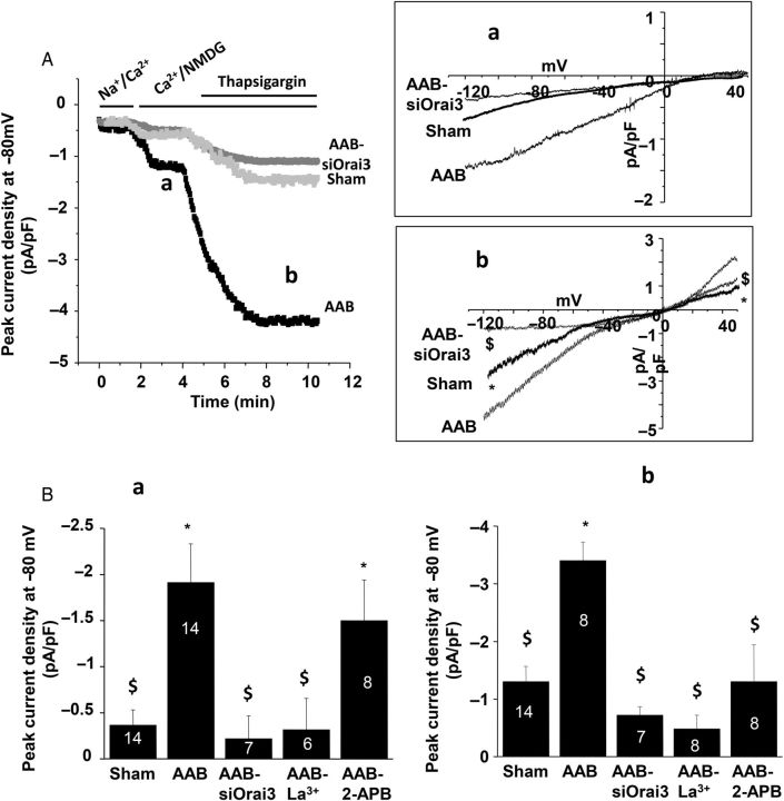
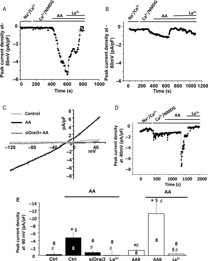
Methods and results: We characterized the expression profile of Orai proteins and their interaction with STIM1 in both normal and hypertrophied adult rat ventricular cardiomyocytes. Orai1 and 3 protein levels were unaltered during the hypertrophic process and both proteins co-immunoprecipitated with STIM1. The level of STIM1 and Orai1 were significantly greater in the macromolecular complex precipitated by the Orai3 antibody in hypertrophied cardiomyocytes. We then used a non-viral method to deliver Cy3-tagged siRNAs in vivo to adult ventricular cardiomyocytes and silence Orai channel candidates. Cardiomyocytes were subsequently isolated then the voltage-independent, i.e. store-independent and store-operated Ca(2+) entries were measured on Fura-2 AM loaded Cy3-labelled and control isolated cardiomyocytes. The whole cell patch-clamp technique was used to measure Orai-mediated currents. Specific Orai1 and Orai3 knockdown established Orai3, but not Orai1, as the critical partner of STIM1 carrying these voltage-independent Ca(2+) entries in the adult hypertrophied cardiomyocytes. Orai3 also drove an arachidonic acid-activated inward current.

Conclusion: Cardiac Orai3 is the essential partner of STIM1 and drives voltage-independent Ca(2+) entries in adult cardiomyocytes. Arachidonic acid-activated currents, which are supported by Orai3, are present in adult cardiomyocytes and increased during hypertrophy.

Keywords: Calcium; Cardiac hypertrophy; Orai; STIM1; SiRNA.


Figures

Figure 1
Figure 1
(A–B) Orai1 and Orai3 are expressed in the normal and hypertrophied rat-ventricular cells. Western blot (A) and quantifications (B) of STIM1, Orai1 and 3 in rat left-ventricular cardiomyocytes, normalized to GAPDH (in arbitrary units, a.u.), n = 7 sham, n = 7 AAB animals. Data are represented as mean ± SEM. Comparison between sham and AAB was performed with the Mann–Whitney U test. (C) STIM1, Orai1, and Orai3 are present in the same macromolecular complex from sham and AAB cardiomyocytes and a large recruitment of Orai3 occurs in AAB cells. Co-immunoprecipitation of Orai1, Orai3, or STIM1 with STIM1, Orai1, and Orai3 in left-ventricular cardiomyocytes derived from sham-operated or AAB rats. Each co-immunoprecipitation was repeated with extracts from three Sham and three AAB rats. AAB values were normalized to Sham for each blot. Below is the quantification of the western blot. Statistical analysis was performed using a one-sample test, testing if the mean of the AAB group differs from 1: *P < 0.05. (D and E) Orai3 compensates for the loss of Orai1. Western blot (D) showing the efficient knockdown of Orai1 and 3 in AAB cardiomyocytes. Histograms in (E) representing relative protein levels normalized to GAPDH (n = 3 animals for each condition). Statistical analysis was performed with the Mann–Whitney U test. Data are presented as mean ± SEM. *P < 0.05 vs. scrambled.
Figure 2
Figure 2
Effect of Orai knockdown on constitutive Ca2+ entry. (A and B) Orai1–3 as well as Orai3 knockdown inhibits basal constitutive Ca2+ entry in left-ventricular cardiomyocytes. Comparison between AAB and AAB siOrai1/2/3 was performed with the Student's t-test; while Kruskal–Wallis one-way ANOVA on ranks followed by Dunn's post hoc tests were used for the multiple comparisons. $P < 0.05 vs. sham and AAB, respectively. (C) Orai1 silencing leads to an increase in basal constitutive Ca2+ entry. Representative recordings of Fura2 emission ratio (ΔF340/F380) in the cardiomyocytes under basal conditions (left panel). Quantification of the amplitude (middle panel) and the rate of rise (right panel) of the Fura-2 signal in the various conditions. Numbers in the columns represent the number of cells analysed from three different rats for each condition. Statistical analysis was performed with Kruskal–Wallis one-way ANOVA on ranks followed by Dunn's post hoc tests. Data are presented as mean ± SEM. $P < 0.05 vs. sham and AAB, respectively, *P < 0.05 vs. sham, #P < 0.05 vs. AAB and sham siOrai1.
Figure 3
Figure 3
Effect of Orai knockdown on the store-dependent Ca2+ entry. (A and B) Orai1–3 as well as Orai3 knockdown inhibits SOCE in left-ventricular cardiomyocytes. Comparison between AAB and AAB siOrai1/2/3 was performed with the Student's t-test; while Kruskal–Wallis one-way ANOVA on ranks followed by Dunn's post hoc tests were used for the multiple comparisons. $P < 0.05 vs. sham Tg and AAB Tg, respectively. (C) Orai1 silencing leads to an increase in SOCE. Representative recordings of Fura2 emission ratio (ΔF340/F380) in the cardiomyocytes under basal conditions (left panel). Quantification of the amplitude (middle panel) and the rate of rise (right panel) of the Fura-2 signal in the various conditions. Numbers in the columns represent the number of cells analysed from three different rats for each condition. Statistical analysis was performed with Kruskal–Wallis one-way ANOVA on ranks followed by Dunn's post hoc tests. Data are presented as mean ± SEM. $P < 0.05 vs. sham Tg and AAB Tg, respectively, *P < 0.05 vs. sham Tg, #P < 0.05 vs. AAB Tg and sham Tg siOrai1.
Figure 4
Figure 4
Orai3-dependent cation currents in adult cardiomyocytes. Whole-cell patch-clamp recordings in ventricular cells. (A) Current density recorded at −80 mV in control cardiomyocytes, hypertrophic cardiomyocytes (AAB), and hypertrophic cardiomyocytes transfected with siOrai3 (AAB+siOrai3). (Aa) Typical current–voltage relationship of a store-independent current revealed by replacing external Na+ with NMDG. (Ab) Typical current–voltage relationship of store-dependent current revealed by thapsigargin application obtained after subtraction of the store-independent current (a). (B) Mean values of store-independent (a) and store-dependent (b) peak current density recorded at −80 mV. Mean values in b were calculated on thapsigargin-inducing current after substraction of the store-independent current. In AAB cardiomyocytes, La3+ (100 µM) or siOrai3 inhibits both the store-independent and store-dependent currents (a), whereas 2-APB addition (10 µM) only affects thapsigargin-induced current (b). Numbers in the columns represent the number of cells isolated from three to five rats. Data are presented as mean ± SEM. *P < 0.05 compared with sham. $P < 0.05 compared with non-transfected AAB cardiomyocytes.
Figure 5
Figure 5
Orai3 carries an arachidonic acid-inducing current in control and AAB cardiomyocytes. (A) Whole-cell patch-clamp recordings at −80 mV in control adult ventricular cells before and after arachidonic acid (AA; 8 µM), followed by La3+ (100 µM) external application. (B). Whole-cell patch-clamp recordings at −80 mV in siOrai3 transfected control adult ventricular cells before and after arachidonic acid (AA; 8 µM), followed by La3+ (100 µM) external application. (C) Typical current–voltage relationship of AA-inducing current obtained in control and transfected adult ventricular cardiomyocytes. Numbers in the columns represent the number of cells isolated from three rats. (D). Whole-cell patch-clamp recordings at −80 mV in adult AAB ventricular cells before and after arachidonic acid (AA; 8 µM), followed by La3+ (100 µM) external application. (E) Mean values of AA-inducing current recorded at −80 mV in the presence of Ca2+ and NMDG. Analysis was performed on eight cardiomyocytes isolated from three control animals (ctrl) and four AAB animals. Data are presented as mean ± SEM. *P < 0.05 compared with control conditions in the absence of AA. $P < 0.05 compared with AAB conditions in the absence of AA. #P < 0.05 ctrl in the presence of AA.

Comment in

References

    1. Putney JW., Jr Identification of cellular activation mechanisms associated with salivary secretion. Annu Rev Physiol. 1986;48:75–88. - PubMed
    1. Parekh AB, Penner R. Store depletion and calcium influx. Physiol Rev. 1997;77:901–930. - PubMed
    1. Hunton DL, Lucchesi PA, Pang Y, Cheng X, Dell'Italia LJ, Marchase RB. Capacitative calcium entry contributes to nuclear factor of activated T-cells nuclear translocation and hypertrophy in cardiomyocytes. J Biol Chem. 2002;277:14266–14273. - PubMed
    1. Hunton DL, Zou L, Pang Y, Marchase RB. Adult rat cardiomyocytes exhibit capacitative calcium entry. Am J Physiol Heart Circ Physiol. 2004;286:H1124–H1132. - PubMed
    1. Collins HE, Zhu-Mauldin X, Marchase RB, Chatham JC. STIM1/Orai1-mediated SOCE: current perspectives and potential roles in cardiac function and pathology. Am J Physiol Heart Circ Physiol. 2013;305:H446–H458. - PMC - PubMed

Publication types

MeSH terms